Hostname: page-component-68c7f8b79f-kbpd8 Total loading time: 0 Render date: 2025-12-23T14:50:07.809Z Has data issue: false hasContentIssue false

Biological properties of vitamins of the B-complex, part 2 – vitamins B6 and B7 (biotin, vitamin H)

Published online by Cambridge University Press:  30 May 2025

Patrícia Dias
Affiliation:
Department of Pharmacology and Toxicology, Faculty of Pharmacy in Hradec Králové, Charles University, Hradec Králové, Czech Republic
Tomáš Siatka
Affiliation:
Department of Pharmacognosy, Faculty of Pharmacy in Hradec Králové, Charles University, Hradec Králové, Czech Republic
Marie Vopršalová
Affiliation:
Department of Pharmacology and Toxicology, Faculty of Pharmacy in Hradec Králové, Charles University, Hradec Králové, Czech Republic
Monika Moravcová
Affiliation:
Department of Pharmacology and Toxicology, Faculty of Pharmacy in Hradec Králové, Charles University, Hradec Králové, Czech Republic
Jana Pourová
Affiliation:
Department of Pharmacology and Toxicology, Faculty of Pharmacy in Hradec Králové, Charles University, Hradec Králové, Czech Republic
Nikola Přívratská
Affiliation:
Department of Clinical Biochemistry and Diagnostics, University Hospital Hradec Králové, Hradec Králové, Czech Republic Department of Analytical Chemistry, Faculty of Pharmacy in Hradec Králové, Charles University, Hradec Králové, Czech Republic
Lenka Kujovská Krčmová
Affiliation:
Department of Clinical Biochemistry and Diagnostics, University Hospital Hradec Králové, Hradec Králové, Czech Republic Department of Analytical Chemistry, Faculty of Pharmacy in Hradec Králové, Charles University, Hradec Králové, Czech Republic
Lenka Javorská
Affiliation:
Department of Clinical Biochemistry and Diagnostics, University Hospital Hradec Králové, Hradec Králové, Czech Republic
Přemysl Mladěnka*
Affiliation:
Department of Pharmacology and Toxicology, Faculty of Pharmacy in Hradec Králové, Charles University, Hradec Králové, Czech Republic
*
Corresponding author: Přemysl Mladěnka; Email: mladenkap@faf.cuni.cz
Rights & Permissions [Opens in a new window]

Abstract

Vitamins B6 (that is, pyridoxin and its analogues) and B7 (that is, biotin or vitamin H) are essential molecules for many physiological processes. In addition to their well-known involvement in several enzymatic reactions, recent discoveries revealed their participation in other processes, for example, in gene expression via epigenetic processes, such as biotinylation of proteins in the case of biotin. Plants, fungi, archaea and most bacteria synthesise both vitamins, whereas animals and humans lack enzymes for their biosynthesis and depend on their exogenous supply. At least in the case of biotin, human gastrointestinal microbiota can likely partly satisfy the need. Both vitamins are water soluble and require a transporter for efficient absorption after oral administration; they can be rapidly excreted; hence, they are considered largely non-toxic. In addition to physiological and kinetic aspects of vitamin B6 and biotin, this review, which is based on a search in PubMed up to 2023, covers sources of these vitamins, the impact of food treatment on their content, causes and symptoms of deficiency and specific mutations related to their function. Currently available literature on the analytical determination of these vitamins in biological fluids, possible pharmacological uses and symptoms of toxicity, although rare, are also included.

Information

Type
Review Article
Creative Commons
Creative Common License - CCCreative Common License - BY
This is an Open Access article, distributed under the terms of the Creative Commons Attribution licence (https://creativecommons.org/licenses/by/4.0/), which permits unrestricted re-use, distribution and reproduction, provided the original article is properly cited.
Copyright
© The Author(s), 2025. Published by Cambridge University Press on behalf of The Nutrition Society

Introduction

Vitamins of the B-complex represent water-soluble molecules with essential roles in humans. The present review is a follow-up to our previous manuscript, in which we summarised the biological properties of the vitamins B1, B2, B3 and B5 (Reference Hrubša, Siatka and Nejmanová1). Herein, we centre on vitamins B6 and B7 (biotin) to provide a comprehensive summary of sources, properties, physiological functions, disorders that result from their deficiency and scientific information, which has been often overlooked since their discovery. We sought to cover all significant studies on the topic, including current trends and potential directions for future research. Such a review has been previously missing in the available literature.

Methods

PubMed was used as the bibliography database, and eligible publications were selected from 1938 to 2023. The following keywords were added to the query box: (vitamin B6 AND properties) and (vitamin B6 AND sources) and (vitamin B6 AND pharmacokinetics), (vitamin B6 AND physiological function), (vitamin B6 AND pharmacological uses), (vitamin B6 AND toxicity). Instead of vitamin B6, similar combinations were used with pyridoxine, vitamin B7 and biotin. The eligibility criteria were as follows: peer-reviewed journal articles or book chapters published in the English language. There were no exclusion criteria for the search.

Vitamin B6

An introduction to vitamin B6

Vitamin B6, ordinarily but imprecisely known as pyridoxine, is a general term for water-soluble pyridine derivatives with the same physiological role. This vitamin comprises six related compounds – vitamers (Fig. 1a), that is, pyridoxine (or pyridoxole, an alcohol), pyridoxal (an aldehyde), pyridoxamine (an amine) and their 5′-phosphate esters, such as pyridoxal 5′-phosphate (PLP), pyridoxamine 5′-phosphate and pyridoxine 5′-phosphate. PLP is the biologically active form of vitamin B6 because it is a cofactor of most vitamin B6-dependent enzymes in the organism(Reference McCormick, Bowman and Russell2,Reference Mooney, Leuendorf, Hendrickson and Hellmann3) .

Fig. 1. Chemical structures of vitamin B6, including its active forms, and vitamin B7. (a) Structure of the vitamers of B6. (b) Vitamin B6 salvage pathway. PK, pyridoxine/pyridoxamine/pyridoxal kinase; PNPO, pyridoxine phosphate oxidase. (c) Chemical structure of D(+)-biotin. The biotin molecule is composed of two rings: an imidazolidinone ring (blue) and a tetrahydrothiophene group (red) attached to a valeric acid moiety as a side chain (yellow).

Pyridoxine was discovered in 1934 by Hungarian physician Paul György and colleagues, and was isolated in pure form shortly thereafter. Humans must acquire it from their diet. Moreover, PLP can be recycled from food and degraded vitamin B6 in the salvage pathway when the vitamin undergoes interconversion inside cells and yields different forms, including active PLP (Fig. 1b)(Reference Eliot and Kirsch4,Reference Leklem5) . Pyridoxine, pyridoxal and pyridoxamine are converted to their phosphorylated forms by the pyridoxine/pyridoxamine/pyridoxal kinase, while phosphatases hydrolyze phosphorylated vitamin B6 vitamers. Pyridoxine 5′-phosphate and pyridoxamine 5′-phosphate are further oxidised to the active form, PLP, by the enzyme pyridoxine (pyridoxamine) phosphate oxidase.

Sources of vitamin B6

Natural sources of vitamin B6

Plants, fungi, archaea and most bacteria synthesise pyridoxine, whereas animals and humans lack enzymes for its biosynthetic pathway and rely solely on the exogenous supply of the vitamin(Reference Mooney, Leuendorf, Hendrickson and Hellmann3,Reference Tanaka, Tateno and Gojobori6Reference Chen and Xiong27) . Vitamin B6 is widely distributed in foods of plant and animal origin. Whole grains, bananas, potatoes, pulses, nuts, beef, pork, poultry, organ meats and fish are good sources for humans(Reference da Silva, Gregory, Marriott, Birt, Stallings and Yates28Reference Planells, Sanchez, Montellano, Mataix and Llopis50). Some herbs and spices (for example, garlic, curry, and ginger)(51), some gluten-free pseudocereals (for example, amaranth)(Reference Rybicka and Gliszczynska-Swiglo52) and royal jelly are also rich in vitamin B6 (Reference McDowell37,Reference Kamyab, Gharachorloo, Honarvar and Ghavami53) . In animal-derived foods, vitamin B6 is usually present in phosphorylated forms (mainly of pyridoxal and pyridoxamine) and, to a lesser extent, in the free form(Reference Liu, Farkas, Wang, Kohli and Fitzpatrick23,Reference McDowell37,Reference Ollilainen54Reference Schmidt, Schreiner and Mayer56) . There is limited information on the bioavailability of vitamin B6 from animal products in humans. The bioavailability is estimated to be generally high and, in many cases, almost complete. However, thermal processing reduces it by 25–30%; and the reaction between pyridoxal and pyridoxal phosphate with the ε-amino group of protein-bound lysine may be responsible for the decreased bioavailability(Reference Reynolds57Reference Kabir, Leklem and Miller61). In plant-derived foods, the vitamin usually occurs as both free pyridoxine and in a glycosylated form, particularly as pyridoxine-β-d-glucoside, whose proportion can range depending on the plant species, from 5% to 75% of the total vitamin content(Reference Liu, Farkas, Wang, Kohli and Fitzpatrick23,Reference da Silva, Gregory, Marriott, Birt, Stallings and Yates28,Reference Ollilainen54,Reference Reynolds57,Reference Gregory62Reference Mangel, Fudge, Gruissem, Fitzpatrick and Vanderschuren68) . The glucoside is only partly cleaved enzymatically by hydrolases in the small intestine, and its bioavailability is about 50% and 75% lower than that of free pyridoxine in humans and rats, respectively, that is to say, that apparently the capability of utilising the glycosylated form is species specific. The contribution of pyridoxine-β-d-glucoside to the total vitamin B6 intake in the average human diet is around 15%, hence different types of vegetarian diet do not pose a risk for vitamin B6 deficiency. This fact is also supported by findings from a population-based survey comparing the vitamin B6 status among vegetarians, pescatarians, flexitarians and meat-eaters. However, individuals with a marginal intake of total vitamin B6 would be more prone to reduced nutritional status due to this incomplete bioavailability(Reference da Silva, Gregory, Marriott, Birt, Stallings and Yates28,29,Reference Waldmann, Dörr, Koschizke, Leitzmann and Hahn46,Reference Ollilainen54,Reference Reynolds57,Reference Ink, Gregory and Sartain58,Reference Gregory62,Reference Gregory64,Reference Gregory, Trumbo, Bailey, Toth, Baumgartner and Cerda69Reference Strain, Hughes, Pentieva, Ward, Hoey, McNulty, Biesalski, Drewnowski, Dwyer, Strain, Weber and Eggersdorfer83) . The absolute bioavailability of vitamin B6 from a mixed diet is estimated to be about 75%(29,Reference Jungert, Linseisen, Wagner and Richter44,Reference Spinneker, Sola, Lemmen, Castillo, Pietrzik and Gonzalez-Gross84Reference Tarr, Tamura and Stokstad86) .

Vitamin B6 is also synthesised in significant quantities by the microbiota of the human large intestine, and this could represent a secondary exogenous source of the vitamin. Indeed, the existence of a specific carrier-mediated mechanism for pyridoxine uptake in human colonocytes was demonstrated. Conversely, it is likely that a large portion of the vitamin produced by microbiota is taken up by non-synthesising microbes. The extent of the contribution of microbially produced vitamin B6 to overall body levels is unclear as there are no human studies to provide evidence for it(Reference Magnusdottir, Ravcheev, de Crecy-Lagard and Thiele22,Reference da Silva, Gregory, Marriott, Birt, Stallings and Yates28,29,Reference McDowell37,Reference Combs and McClung87Reference Rodionov, Arzamasov and Khoroshkin93) . Amounts of vitamin B6 in some selected foodstuffs are shown in Table 1.

Table 1. Vitamin B6 content in selected foodstuffs

Antivitamins B6

The diet can also contain antivitamin B6 that either compete for reactive sites of vitamin B6-requiring enzymes or directly inactivate the vitamin(Reference McDowell37,Reference Mayengbam, House and Aliani94) . The best-known antivitamin B6 is probably ginkgotoxin (4’-O-methylpyridoxine), which occurs in different tissues of the tree Ginkgo biloba, with the highest concentrations being present in seeds. Ingestion of ginkgotoxin can lead to abdominal pain, epileptiform convulsions and loss of consciousness due to the aforementioned interference with vitamin B6. As seeds are a food source in Southeast Asia, including China, Japan and Korea, and extracts from leaves are used in pharmaceutical products worldwide, they represent a potential health risk(Reference Mooney, Leuendorf, Hendrickson and Hellmann3,Reference Wada, Ishigaki, Ueda, Sakata and Haga95Reference Gong, Wu, Fan, Li, Wang and Wang112) . Indeed, ginkgotoxin and its derivatives found in the African trees of the genus Albizia (for example, A. tanganyicensis, A. versicolor, A. julibrissin and A. lucida) are the cause of poisoning of livestock (cattle and sheep): one of the most important agricultural problems in South Africa(Reference Mooney, Leuendorf, Hendrickson and Hellmann3,Reference Leistner and Drewke101,Reference Steyn, Vleggaar and Anderson113) . Flaxseed contains the vitamin B6 antagonists, 1-amino-d-proline, and its precursor, the dipeptide linatine. Their possible deleterious effects through the consumption of flaxseed deserve attention in individuals with moderate vitamin B6 status(Reference Mayengbam, House and Aliani94,Reference Mayengbam, Raposo, Aliani and House114Reference Klosterman118) . Gyromitrin (N-methyl-N-formylhydrazone) from the toxic mushroom Gyromitra esculenta (genus Gyromitra is also known as false morrel) is converted to (mono)methylhydrazine after ingestion, which is able to inhibit pyridoxal kinase and hence depletes vitamin B6. Intoxication usually occurs about 10 h after the ingestion of fresh or dried mushrooms. It gives rise to poisoning symptoms such as confusion and seizures. Interestingly, during cooking, methylhydrazine volatilises, and poisoning occurs also after inhalation of these vapours(Reference Klosterman118Reference Horowitz, Kong and Horowitz122). Similarly, agaritine containing a hydrazinic moiety in its structure is a toxic principle of various Agaricus species, for example, the edible button mushroom Agaricus bisporus (Reference Klosterman118,Reference Lagrange and Vernoux121,Reference Berdanier, Berdanier, Dwyer and Feldman123) . The content of both toxins in fungi may be decreased by processing, such as boiling in water, drying and freezing(Reference Lagrange and Vernoux121,Reference Arshadi, Nilsson and Magnusson124,Reference Roupas, Keogh, Noakes, Margetts and Taylor125) . Other natural vitamin B6 antagonists, which are of little significance to human nutrition, are toxic non-proteinogenic amino acids occurring in some leguminous plants: mimosine in Mimosa and Leucaena species, and canavanine and canaline in Canavalia species(Reference Klosterman118,Reference Klosterman126Reference Gregory and Kirk132) .

Effects of food processing on vitamin B6 content

Food processing is the transformation of agricultural products into foods for human consumption. Primary processing is the conversion of the inedible raw products into food ingredients. Secondary processing involves the conversion of food ingredients into edible foods. Tertiary processed foods are commercially prepared foods. Products from primary processes make up the major part of the human diet as they are either consumed raw or used as ingredients in secondary and tertiary processes(Reference Özilgen, Özilgen and Hitzmann133). Food processing may alter the vitamin B6 content(Reference Berry Ottaway, Skibsted, Risbo and Andersen134,Reference Godoy, Amaya-Farfan, Rodriguez-Amaya, Rodriguez-Amaya and Amaya-Farfan135) . A rough overview of the major data on vitamin B6 losses in some food groups due to processing is presented in Supplementary Table S1 in the Supplementary Data. More data on specific foods, information on conditions and comments are in the text below.

Milling and refining of cereals

The primary processing of cereals (milling and refining) that separates the bran and germ, which are rich in micronutrients, from starchy endosperm causes a considerable loss of vitamin B6 (Reference Slavin, Jacobs and Marquart136Reference Brouns, Hemery, Price and Anson141). Milling reduces the value of the vitamin B6 content in maize by 65–75%(Reference Thielecke, Lecerf and Nugent137,Reference Titcomb and Tanumihardjo142Reference Dunn, Jain and Klein146) . The vitamin B6 content decreases by 66–89% in white wheat flour, compared with wholegrain flour(Reference Shewry and Hey45,Reference Slavin, Jacobs and Marquart136Reference Călinoiu and Vodnar138,Reference Titcomb and Tanumihardjo142Reference Hegedüs, Pedersen and Eggum144,Reference Henry and Heppell147) . The content of vitamin B6 is likewise 64% and 79.5% lower in refined than in wholegrain rye and sorghum flour, respectively.(Reference Hegedüs, Pedersen and Eggum144). Vitamin B6 losses in non-parboiled and parboiled white rice are 42–86% and 12–26%, respectively, compared with brown rice. The decline in vitamin B6 in parboiled rice is lower, in contrast to the non-parboiled one, because a part of the vitamin diffuses from the vitamin-rich outer bran layer into the endosperm during the parboiling process that takes place before milling(Reference Mangel, Fudge, Gruissem, Fitzpatrick and Vanderschuren68,Reference Thielecke, Lecerf and Nugent137,Reference Titcomb and Tanumihardjo142,Reference Garg, Sharma and Vats143,Reference Tiozon, Fernie and Sreenivasulu148Reference Schroeder152) . The secondary processing of cereals, such as breadmaking, rice cooking and nixtamalisation of maize, brings on additional vitamin B6 losses. They are discussed later (‘Processing of plant-based foods’).

Properties of vitamin B6 and mechanisms of vitamin loss during food processing

Vitamin B6 loss during processing and storage of food can occur in several ways. Being soluble in water, leaching is one of the principal causes. Vitamin B6 in foods is stable under acidic conditions but unstable in neutral and alkaline environments, particularly when exposed to heat or light. The acidic aqueous solutions of vitamin B6 may be heated without decomposition, as vitamin B6 is destroyed by ultraviolet radiation in neutral or alkaline solutions but not in acidic solutions. Vitamin B6 is normally stable to oxygen. Of the several vitamers, pyridoxine is far more stable than pyridoxal and pyridoxamine. Therefore, the processing losses of vitamin B6 tend to be highly variable, with plant-derived foods (containing mostly pyridoxine) losing little of the vitamin, and animal products (containing mostly pyridoxal and pyridoxamine) associated with higher losses(Reference McDowell37,Reference Gregory, Ink and Sartain59,Reference Combs and McClung87,Reference Gregory and Kirk132,Reference Berry Ottaway, Skibsted, Risbo and Andersen134,Reference Godoy, Amaya-Farfan, Rodriguez-Amaya, Rodriguez-Amaya and Amaya-Farfan135,Reference Henry and Heppell147,Reference Aboul-Enein and Loutfy153Reference Ribeiro, Pinto, Lima, Volpato, Cabral and de Sousa166) .

Processing of animal-based foods

Boiling, stewing, roasting and frying reduce the vitamin B6 content by 55%, 33–58%, 30% and 40–45%, respectively, in pork; by 60–77%, 55–57%, 40% and 55–58%, respectively, in beef; and by 40–58%, 40–47%, 50% and 45–56%, respectively, in chicken, depending on cooking temperature and time(Reference Shibata, Yasuhara and Yasuda161,Reference Mašková, Rysova, Fiedlerova and Holasova167Reference Çatak and Çaman171) . In whole meat dishes, including cooking liquid, gravy, juice or soup, about 15–20% more vitamin B6 remains, owing to retention of the vitamin that leached into the water phase(Reference Bognár168,Reference Bognár170,172Reference Meyer, Mysinger and Wodarski174) . Fried breaded meats contain 5–35% more vitamin B6 than those without breading, which may assist in trapping the liquid and, therefore, decreasing the loss of water-soluble vitamins(Reference Bognár170,Reference Olds, Vanderslice and Brochetti175) . About 9% of vitamin B6 was lost from pork and beef when the drip exuding from the frozen meat during thawing was discarded(Reference Pearson, Burnside, Edwards, Glasscock, Cunha and Novak176,Reference Pearson, West and Luecke177) . The cooking loss of vitamin B6 in fish meat (gilthead seabream, anchovy and Atlantic bonito) was 55–85% and 60–89% when grilled and baked, respectively, due to thermal degradation and leakage of the vitamin in the lost water(Reference Çatak, Çaman and Ceylan178). Heat-induced reduction of vitamin B6 in milk is usually 5–20%, 5–10%, 5–20%, 10–50% and 40% for boiled, pasteurised, ultra-high temperature treated, sterilised and condensed milk, respectively, compared with raw milk(Reference Berry Ottaway, Skibsted, Risbo and Andersen134,Reference Bognár170,Reference Muehlhoff, Bennett and McMahon179Reference Amador-Espejo, Gallardo-Chacon, Nykänen, Juan and Trujillo184) . Hard cooked, poached, scrambled, baked and fried eggs lose 20–23%, 15%, 10%, 10% and 10% of vitamin B6 during cooking, respectively(Reference Bognár170,Reference Roe, Church, Pinchen and Finglas185,Reference Öhrvik, Carlsen, Källman and Martinsen186) .

Processing of plant-based foods

Boiling, steaming and frying lead usually to a vitamin B6 loss of 30–35%, 15% and 10%, respectively, in vegetables alone, and to that of about 10% when taking the total dish into account(Reference Bognár168,Reference Bognár170) . In chickpeas, microwave cooking, autoclaving and boiling caused a decline of 19%, 34% and 42% in vitamin B6 content, respectively(Reference Alajaji and El-Adawy187). The amount of vitamin B6 in potatoes is reduced by 30–57%, 21% and 10% during boiling, baking and deep frying, respectively(Reference Shibata, Yasuhara and Yasuda161,Reference Bognár169,Reference Roe, Church, Pinchen and Finglas188) . The way of cooking rice influences the content of vitamin B6. In different rice varieties, the boiling cooking method (cooking rice with extra water and then eliminating the water) led to vitamin losses of 3–74%, compared with the traditional cooking method (cooking with a constant amount of water without removing the water)(Reference Rezaei, Alizadeh Sani and Amini189). During breadmaking, the vitamin B6 content decreased on average by 33% and 62% in whole and white wheat bread, respectively, in comparison with whole and white wheat flour(Reference Batifoulier, Verny, Chanliaud, Rémésy and Demigné190,Reference Nurit, Lyan, Pujos-Guillot, Branlard and Piquet191) . Similar results were obtained during rye sourdough bread production(Reference Mihhalevski, Nisamedtinov, Hälvin, Ošeka and Paalme192). Toasting wheat bread induced an increase in vitamin B6 by 75% due to its release from glycosidic bound forms(Reference Nurit, Lyan, Pujos-Guillot, Branlard and Piquet191). Effects of extrusion techniques on vitamin B6 retention in cereal grains showed a reduction of 0–23% and of 65% in maize grits and oat whole grains, respectively(Reference Athar, Hardacre, Taylor, Clark, Harding and McLaughlin193). Drying of tarhana, a traditional Turkish fermented cereal food, resulted in vitamin B6 losses of 3%, 16% and 23% at temperatures of 50 °C, 60 °C and 70 °C, respectively(Reference Ekıncı194). A decrease in vitamin B6 content in nuts varied from 2–7.5% in almonds, up to 4–34% in pistachio nuts after roasting(Reference Bulló, Juanola-Falgarona, Hernández-Alonso and Salas-Salvadó195,Reference Stuetz, Schlörmann and Glei196) . Alkali-processing of corn grains to masa (nixtamalisation) resulted in a loss of 23% of vitamin B6 (Reference Gwirtz and Garcia-Casal145). The highly variable content of vitamin B6 in beer is affected by several factors, including raw materials and the brewing process(Reference Romanini, Rastelli, Donadini, Lambri and Bertuzzi197Reference Bertuzzi, Mulazzi, Rastelli, Donadini, Rossi and Spigno199). Germination is an effective way to improve the nutrition value of edible seeds: increases of 54%, 78% and 26% in vitamin B6 content occurred in germinated lentils(Reference Zhang, De Silva, Dissanayaka, Warkentin and Vandenberg200), rough rice(Reference Moongngarm and Saetung201) and faba beans(Reference Khalil and Mansour202), respectively. Conversely, vitamin B6 levels decreased by 11%, 13% and 50% in germinated wheat(Reference Žilić, Basić, Hadži-Tašković Šukalović, Maksimović, Janković and Filipović203), brown rice(Reference Moongngarm and Saetung201) and sorghum(Reference Pinheiro, Anunciação and de Morais Cardoso204), respectively, after germination.

Food preservation and storage

Canning, a food conservation method, brought on a vitamin B6 reduction of 46%, 34%, 31% and 18% in mushrooms, whole peeled tomatoes, white asparagus and lentils compared with their respective unprocessed products(Reference Martin-Belloso and Llanos-Barriobero205). Ionising irradiation, a method used for food preservation, has a low effect on vitamin B6; losses ranging from zero in wheat to about 15% in fish were observed(Reference Kilcast206,Reference Woodside207) .

The amount of vitamin B6 in button mushrooms significantly declined by 23% and 45% after 6 and 12 months, respectively, during frozen storage at −20 °C(Reference Bernaś and Jaworska208). The content of vitamin B6 decreased gradually in aseptically packaged ultra-high temperature treated milk during storage at room temperature, resulting in a 96% loss after 20 weeks(Reference Oamen, Hansen and Swartzel183). No remarkable changes and a 20% decline in vitamin B6 content happened in vacuum-packaged broccoli au gratin and salmon, respectively, stored at room temperature, either on the Earth or exposed to spaceflight for 880 d; the vitamin content in flight samples did not degrade faster than that of ground controls(Reference Zwart, Kloeris, Perchonok, Braby and Smith209). The investigation of the influence of storage conditions on vitamin B6 retention in a freeze-dried tuna mornay meal (containing tuna, vegetables and pasta) fortified with that vitamin showed a mean decrease of 14% in the vitamin following storage at temperatures of 1 °C, 30 °C and 40 °C for up to 24 months(Reference Coad and Bui210). The vitamin B6 losses in meals in two hospital foodservice systems, the cook/hot-hold system, where food is held hot from the time of cooking to service, and the cook/chill system, where the cooked food is chilled, stored and reheated, have also been summarised and compared(Reference Williams211).

Industrial production of vitamin B6

Pyridoxine hydrochloride, which is mainly used in pharmaceutical preparations, dietary supplements and as an additive in food and feed, is manufactured by chemical synthesis(29,Reference McDowell37,60,Reference Spinneker, Sola, Lemmen, Castillo, Pietrzik and Gonzalez-Gross84,Reference Combs and McClung87,Reference Bonrath, Zhang, Pauling, Weimann, Bellussi, Bohnet, Bus, Drauz, Greim, Jackel, Karst, Kleemann, Kreysa, Laird, Meier, Ottow, Roper, Scholtz, Sundmacher, Ulber and Wietelmann158,Reference Fischesser, Fritsch, Gum, Karge and Keuper212Reference Gum219) . All present-day industrial vitamin B6 syntheses use the Diels–Alder reaction of a diene (4,5-substituted oxazoles) and a dienophile (alkyldioxepins) as a key step(Reference Bonrath, Zhang, Pauling, Weimann, Bellussi, Bohnet, Bus, Drauz, Greim, Jackel, Karst, Kleemann, Kreysa, Laird, Meier, Ottow, Roper, Scholtz, Sundmacher, Ulber and Wietelmann158,Reference Eggersdorfer, Laudert and Letinois220Reference Ledesma-Amaro, Jiménez, Revuelta, McNeil, Archer, Giavasis and Harvey225) . An alternative to the current chemical processes might be environmentally sustainable bioprocesses based on microbial vitamin B6 fermentation, which is of great interest to the biotechnological industry. Several attempts have been made to construct overproducing strains by genetic engineering of microorganisms such as Sinorhizobium meliloti, E. coli and Bacillus subtilis. Unfortunately, production levels are too low and are not cost effective. Therefore, major metabolic engineering efforts are still required for developing fermentation processes that could outcompete the chemical synthesis of vitamin B6. The main bottlenecks are insufficient activities of some enzymes in the biosynthetic pathway and accumulation of toxic intermediate metabolites(Reference Acevedo-Rocha, Gronenberg, Mack, Commichau and Genee226Reference Richts and Commichau237).

Food fortification and biofortification with vitamin B6

Food fortification is defined as the practice of deliberately adding an essential micronutrient to food that is commonly consumed by the general population with the intention of improving the nutritional quality of the food supply and providing a public health benefit with minimal risk to health(Reference Cardoso, Fernandes, Gonzaléz-Paramás, Barros and Ferreira238Reference Allen, De Benoist, Dary and Hurrell240). Foods fortified with vitamin B6, similarly to dietary supplements, constitute an additional dietary source of the vitamin(60,Reference de Pee150,Reference Bird, Murphy, Ciappio and McBurney241Reference Ho, Quay, Devlin and Lamers246) . Overall, vitamin B6 deficiency is rare in the general healthy population(Reference Parra, Stahl and Hellmann8,29,Reference Jungert, Linseisen, Wagner and Richter44,Reference de Pee150,Reference Tucker, Olson, Bakun, Dallal, Selhub and Rosenberg243,Reference Ho, Quay, Devlin and Lamers246Reference Brown, Ameer and Daley250) . It may be a concern in high-income as well as low-income countries in certain groups(Reference Titcomb and Tanumihardjo142), such as older adults(Reference Tang, Xu and Shiu-Ming245,Reference Morris, Picciano, Jacques and Selhub251Reference ter Borg, Verlaan and Hemsworth254) , people of low socio-economic status and those experiencing food insecurity(Reference Titcomb and Tanumihardjo142,Reference Bird, Murphy, Ciappio and McBurney241,Reference Duvenage and Schönfeldt244,Reference Tang, Xu and Shiu-Ming245,Reference Brown, Ameer and Daley250) . As for 2022, some countries, mostly but not solely located in Africa, have mandatory fortification of wheat flour (most often), maize flour and/or rice with vitamin B6 (Nicaragua, Panama, Cuba, Peru, Jordan, Palestine, Nigeria, Chad, Ethiopia, Kenya, Uganda, Rwanda, Burundi, Tanzania, Mozambique, Zimbabwe and South Africa)(255Reference Swanepoel, Havemann-Nel and Rothman257). There is a voluntary fortification with vitamin B6 in many other countries, such as the USA, the Dominican Republic, Eswatini, India, Bangladesh, Myanmar, the UK and countries of the European Union; the vitamin is added to various foods, such as atta, maida, rice, breakfast cereals, beverages and cereal-based foods for infants and young children(Reference da Silva, Gregory, Marriott, Birt, Stallings and Yates28,60,Reference Titcomb and Tanumihardjo142,Reference de Pee150,Reference Berendsen, van Lieshout, van den Heuvel, Matthys, Péter and de Groot217,255,256,258Reference Hannon, Kiely and Flynn265) .

Biofortification is a process of increasing the density of micronutrients (vitamins and minerals) in a crop and comprises (sensu stricto, that is, omitting agronomic practices) conventional plant breeding and genetic engineering approaches. It differs from fortification because it aims to make plant foods naturally more nutritive rather than adding nutrients to the foods during food processing. Biofortification is an ideal strategy to improve nutrition for rural and poor communities that rely on subsistence farming for nutrition or may not have access to diverse diets, supplements and fortified foods. Biofortification complements existing interventions and may help by increasing the daily adequacy of micronutrient intake among the most vulnerable micronutrient deficient people(Reference Titcomb and Tanumihardjo142,Reference Malik and Maqbool239,Reference Bouis266Reference Strobbe and Van Der Straeten268) . Vitamin B6 is de novo synthesised by plants, and therefore, biofortification could be a promising route to enhance food quality by increasing the vitamin levels in plants in the future(Reference Parra, Stahl and Hellmann8,Reference Vanderschuren, Boycheva, Li, Szydlowski, Gruissem and Fitzpatrick269Reference Blancquaert, De Steur, Gellynck and Van Der Straeten271) . Analysis of the natural diversity of vitamin B6 content in wheat, rice and potato germplasm has shown limited variation, so breeding strategies do not seem to be adequate to increase the vitamin content in those crops(Reference Mangel, Fudge, Gruissem, Fitzpatrick and Vanderschuren68,Reference Titcomb and Tanumihardjo142,Reference Shewry, Van Schaik and Ravel272,Reference Mooney, Chen, Kühn, Navarre, Knowles and Hellmann273) , in contrast to maize, where remarkable wide ranges in vitamin B6 levels among various genotypes were recently reported(Reference Azam, Lian, Liang, Wang, Zhang and Jiang274). Most efforts to date have used genetic engineering approaches. Biosynthesis of vitamin B6 is primarily controlled by two enzymes, making vitamin B6 biofortification an attractive target for plant geneticists. Overexpression of genes encoding one or both enzymes leads to the enhanced accumulation of vitamin B6 in transgenic plants compared with the untransformed ones: 0·86–1·25-fold in tobacco plants, 1·45–4-fold in Arabidopsis seeds, 0·16–34·96-fold in wheat seeds, 1·6–3·9-fold in rice seeds, 3–16-fold in cassava roots and 1·07–1·5-fold in potato tubers. Interestingly, enhancing vitamin B6 levels in plants may also positively affect their tolerance to environmental stress(Reference Chen and Xiong27,Reference Titcomb and Tanumihardjo142,Reference Strobbe and Van Der Straeten268Reference Fudge, Mangel, Gruissem, Vanderschuren and Fitzpatrick270,Reference Herrero and Daub275Reference Bagri, Upadhyaya, Kumar and Upadhyaya280) . All biofortification attempts revealed the feasibility of raising the vitamin B6 amounts in plants. So far, the vitamin B6 contents in transgenic plants are low and highly variable. Regardless, more research for understanding the regulatory mechanisms that control genes involved in the biosynthesis and metabolism of vitamin B6 in plants is needed(Reference Ledesma-Amaro, Jiménez, Revuelta, McNeil, Archer, Giavasis and Harvey225,Reference Vanderschuren, Boycheva, Li, Szydlowski, Gruissem and Fitzpatrick269) .

Pharmacokinetics of vitamin B6

The total content of vitamin B6 in the adult human body is about 170 mg(Reference Coburn, Lewis, Fink, Mahuren, Schaltenbrand and Costill281). B6 vitamers are absorbed in the upper small intestine (jejunum) from diet and/or oral supplements. In addition to the dietary sources of the vitamin, humans might also receive vitamin B6 from bacterial microbiota in the large intestine as mentioned earlier(Reference Said88,Reference Hamm, Mehansho and Henderson282,Reference Said283) . All vitamin B6 analogues, that is, pyridoxine, pyridoxamine, and pyridoxal, are present in the diet. Phosphorylated forms undergo dephosphorylation by means of phosphatases prior to absorption into epithelial cells and prior to release into the portal system. Phosphorylated forms are poorly diffusible and, in fact, they are trapped in cells and a dephosphorylation step is necessary for their efflux. The bioavailability of vitamin B6 from supplements is about 95%, whereas the bioavailability of pyridoxin, pyridoxal and pyridoxamine is similar. The presence of fibre in plant sources reduces bioavailability by 5–10%, while the presence of pyridoxine glucoside reduces bioavailability by 75–80%. On average, the bioavailability of vitamin B6 from a mixed diet can be estimated to be about 75%. In fact, absorption in the intestine is mediated both via passive diffusion (that is, a large amount is readily absorbable without cell saturation) and a carrier mediated mechanism (that is, a saturable mechanism). In humans, there is carrier-mediated transport of B6 vitamers via the vitamin B1 (thiamine) transporters THTR1 and THTR2, which belong to the SLC19A2 and SLC19A3 families(Reference Yamashiro, Yasujima, Said and Yuasa284). The maximum concentration (C max) of pyridoxine is usually achieved within 5·5 h(Reference Surtees, Mills and Clayton285,286) . In the liver, all forms of dephosphorylated vitamin B6 are rephosphorylated and finally converted to pyridoxal 5′-phosphate in hepatocytes. Several enzymes, such as ATP-dependent pyridoxine/pyridoxamine/pyridoxal kinase, phosphatases and flavin mononucleotide–dependent pyridoxine phosphate oxidase (PNPO) are involved in these reactions. PNPO converts pyridoxine 5′-phosphate (PNP) and pyridoxamine 5′-phosphate (PMP) into pyridoxal 5′-phosphate (PLP) (Fig. 1b).

Pyridoxal phosphate further binds to albumin in the liver, and it is released into the circulation, where it forms approximately 60% of total circulating B6, with lesser amounts of all three dephosphorylated forms. After dissociation from albumin and dephosphorylation by alkaline phosphatase, free pyridoxal is taken up by erythrocytes and then trapped inside cells in the form of PLP(Reference Anderson, Fulford-Jones, Child, Beard and Bateman287Reference Merrill, Henderson, Wang, McDonald and Millikan292).

Plasma PLP is the most common parameter for determination of vitamin B6 status. Its usual concentration is more than 30 nM in adults(Reference Leklem5). PLP is utilised as a cofactor of many enzymes related to a row of metabolic pathways(Reference Di Salvo, Contestabile and Safo293,Reference Lumeng and Li294) , as will be discussed later. Circulatory PLP passes into breast milk, and also crosses physiological barriers such as the placental and blood–brain barriers. The same mechanism, as in other organs, is described for brain entry and storage, that is, initial dephosphorylation in the blood–brain barrier by means of tissue non-specific alkaline phosphatase (TNSALP), followed by uptake and entrapping of the vitamin in neurons after phosphorylation to PLP(Reference Surtees, Mills and Clayton295).

The major inactive metabolite of PLP is 4-pyridoxic acid. It is formed in the liver and excreted in the urine (Fig. 2). Urinary excretion of this metabolite greater than 3 mmol/d can be used as a marker of adequate short-term vitamin B6 status. Its half-life appears to be 15–20 d(Reference Stanulović, Jeremić, Leskovac and Chaykin296).

Fig. 2. Pharmacokinetics of vitamin B6. The figure summarises the pharmacokinetics of vitamin B6 in the human body. PN, pyridoxine; PNP, pyridoxine 5′-phosphate; PL, pyridoxal; PLP, pyridoxal 5′-phosphate; PM, pyridoxamine; PMP, pyridoxamine 5′-phosphate; TNSALP, tissue non-specific alkaline phosphatase; BB, blood–brain barrier.

There is not a large storage of vitamin B6 in tissues, probably owing to the fact that humans require only small amounts of vitamin B6 from food sources, since the biologically active form, PLP, can be formed not only by interconversion from different B6 vitamers but also using the cofactors from degraded enzymes in the salvage pathway.

Physiological function of vitamin B6

The active form of vitamin B6, PLP, acts as a coenzyme in more than 140 different enzymatic reactions necessary for vital cellular processes(Reference Parra, Stahl and Hellmann8). This function is enabled by the highly reactive aldehyde group of PLP, that forms Schiff bases with the ε amino groups of lysine residues at the active centres of PLP-dependent enzymes. Conversely, binding to lysine residues on some hormonal receptors is responsible for transcriptional modulation. Moreover, the aldehyde group can react with other amino acids in proteins, especially with cysteine or histidine(Reference Phillips297).

PLP is involved in various pathways, such as:

Besides the role of PLP as a cofactor in biochemical reactions, vitamin B6 also plays other important roles in non-enzymatic functions, for example:

Vitamin B6 deficiency and related disorders

Severe vitamin B6 deficiency resulting from inadequate intake (especially from dietary deficit) is rare in the healthy general population. Hypovitaminosis is usually found in association with other B vitamin deficiencies, such as those of folic acid (vitamin B9) and vitamin B12. As aforementioned, it should be emphasised that dietary vitamin B6 deficiency can occur in elderly people (aged 65 years and over)(Reference Bates, Pentieva and Prentice323). Secondary vitamin B6 deficiency is mostly a result of genetic disorders or drug interactions(Reference Plecko and Stöckler324,Reference Wilson, Plecko, Mills and Clayton325) .

Owing to the involvement of vitamin B6 in many metabolic pathways, a lack of sufficient amounts of vitamin B6 vitamers causes various biochemical changes and may lead to significant health problems. In particular, PLP is essential in the synthesis and metabolism of amino acids and neurotransmitters. Loss of function of the PLP-dependent enzyme glutamate decarboxylase leads to decreased levels of the inhibitory neurotransmitter GABA.

Vitamin B6 deficiency in humans is associated with seborrheic dermatitis and cheilosis (including cracks at the corners of the mouth), glossitis with ulceration, anaemia, sensory polyneuropathy, depression, decreased immune function and increased risk of cardiovascular diseases. In children, characteristic symptoms of deficiency are abnormalities in hearing and seizures.(Reference Spinneker, Sola, Lemmen, Castillo, Pietrzik and Gonzalez-Gross84) Seizures are the results of an imbalance between excitatory (glutamate) and inhibitory (GABA) neurotransmitters(Reference Gospe, Olin and Keen326Reference Kurlemann, Ziegler, Grüneberg, Bömelburg, Ullrich and Palm328).

In the population, there are certain groups of people at increased risk of vitamin B6 inadequacy. People with impaired absorption, especially due to malabsorption syndromes (usually associated with Crohn’s disease and ulcerative colitis) and after bariatric surgery, have low vitamin B6 levels. Patients with renal disease, predominantly with chronic renal insufficiency undergoing dialysis, and liver disease tend to have low plasma PLP concentrations. Also, alcoholics need vitamin B6 supplementation because alcohol is metabolised to acetaldehyde, which decreases PLP formation in cells and competes with PLP for protein binding. Additional groups at risk of vitamin inadequacy despite adequate dietary intakes are not solely elderly persons but also those with autoimmune disorders (for example, rheumatoid arthritis), those who are obese and in pregnancy, or those who are taking oral contraceptives(Reference Chiang, Selhub, Bagley, Dallal and Roubenoff329Reference Salam, Zuberi and Bhutta332). Analytical methods for the detection of vitamin B6 are summarised in Table 2. More details are shown in Supplementary Data Table S2, which evaluates individual specific methodologies with the relevant citations from which the information was obtained.

Table 2. Summary of analytical methods for the assessment of vitamins B6 and B7 in biological fluids

B6, pyridoxine; B6-PL, pyridoxal; B6-P5P, pyridoxal-5-phosphate; B6-PM, pyridoxamine; B6-PA, pyridoxic acid; B7, biotin; B9, folic acid; ATB, antibiotic; CL, chemiluminescence; ECD, electrochemical detection; ELISA, enzyme-linked immunosorbent assay; FLD, fluorescence detection; HPLC, high performance liquid chromatography; IS, internal standard; LC-MS, coupling of liquid chromatography and mass spectrometry; MLC, micellar liquid chromatography; MRM, multiple reaction monitoring; MS, mass spectrometer; MS/MS, tandem mass spectrometry; PDA, photodiode array detection; SIM, selected ion monitoring; 2D-LC, two-dimensional liquid chromatography.

Pyridoxine-dependent epilepsy

Pyridoxine-dependent epilepsy (pyridoxine-dependent seizures, vitamin B6-responsive epilepsy) is a rare inherited metabolic disease characterised by recurrent seizures with their onset usually in prenatal, neonatal and postnatal periods or in childhood. Seizures are caused primarily by low levels of GABA due to PLP deficiency, nevertheless, other abnormalities are involved, for example, low levels of adenosine and methionine cycle defects. This type of epilepsy responds to high intravenous doses of vitamin B6, either as pyridoxine or as its active form PLP, but are resistant to conventional antiepileptic drugs(Reference Pena, MacKenzie and Van Karnebeek333). Decreased PLP availability in this disease is caused by mutations in some genes involved in vitamin B6 metabolism, for example:

These metabolic diseases associated with defects in vitamin B6 are summarised in Table 3.

Table 3. Inborn metabolic disorders related to pyridoxine dependent seizures

Clinically used drugs as antivitamins for B6

In addition to natural antivitamins for B6, there are also certain clinically used drugs that have the same effect. Drugs such as theophylline (a bronchodilator used in the treatment of respiratory diseases, for example, asthma) and caffeine (psychostimulant) directly inhibit pyridoxal kinase, the enzyme involved in activation of PLP. In the case of caffeine, such effects are probable solely in intoxication. The result is a PLP deficiency with accompanying reduction in PLP-dependent enzyme activities. Known consequences include neurotoxic reactions, for example, peripheral neuropathy, restlessness, agitation, tremors and seizures(Reference Glenn, Krober, Kelly, McCarty and Weir348Reference Weir, Keniston, Enriquez and McNamee350). It should be mentioned that standardised extracts from Ginkgo biloba are easily available and used in the therapy of a number of conditions, such as peripheral circulatory disturbances, dizziness and tinnitus, etc.(Reference Azuma, Nokura, Kako, Kobayashi, Yoshimura and Wada98,Reference Gandhi, Desai and Ghatge351,Reference Jang, Roh, Jeong, Kim and Sunwoo352) . Hydrazine derivatives, beyond the aforementioned gyromitrin, also include the antituberculosis drug isoniazid (isonicotinic acid hydrazide). Administration of this drug, particularly in overdose, results in not only the inhibition of pyridoxal kinase by the isoniazid metabolite (hydrazone) but also the inactivation of PLP by other isoniazid metabolites (hydrazines and hydrazides), that form, for example, isonicotinilhydrazide, a compound that is easily excreted in the urine(Reference Kook, Cho, Lee, Lee and Lee353). Another antituberculosis drug, cycloserine, reacts with PLP forming covalent complexes that might inhibit pyridoxal kinase(Reference Court, Centner and Chirehwa354). Another group of drugs, including penicillamine and levodopa, form complexes with PLP, but they do not inhibit pyridoxal kinase(Reference Leon, Spiegel, Thomas and Abrams355,Reference Smith and Gallagher356) . In addition, antiepileptic drugs (phenytoin, valproic acid, and carbamazepine) increase the metabolism of vitamin B6 vitamers, resulting in low PLP plasma levels(Reference Linnebank, Moskau and Semmler357).

Dietary recommendation and pharmacological use of vitamin B6

Vitamin B6 is available in both multivitamin preparations with other B vitamins and as a single vitamin preparation. Oral tablets or solutions for parenteral (intravenous, intramuscular) administration are the most common forms; they usually contain pyridoxine hydrochloride or sometimes PLP.

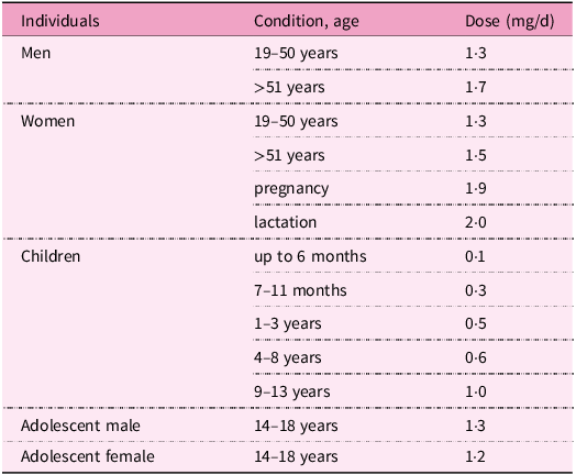
In adults, the current recommended dietary allowances range between 1·3–2·0 mg/d. During pregnancy, lactation and in the elderly, this requirement is increased(286). Recommendations for pyridoxine intake according to age and gender are listed in Table 4.

Table 4. Recommendations for vitamin B6 intake by gender and age(286)

As a supplement, vitamin B6 is used especially in cases of its deficiency, which may be due to insufficient intake or increased need, as specified earlier. As a medication, pyridoxine or PLP are given prophylactically or therapeutically to patients with pyridoxine-dependent epilepsy. In newborns with hereditary syndrome, it is necessary to administer this vitamin in the first week of life to prevent mental retardation or anaemia, and lifelong therapy is necessary. In the literature, however, there is a lack of congruence regarding dose recommendations. The optimal dosage should ensure the control of epileptic seizures, and, at the same time, the absence of side effects in a particular patient. In fact, adequate dosage of pyridoxine requires an individualised regimen according to the desired goal of therapy and tolerance of adverse effects.

Higher doses of pyridoxine are initially administered, for example, in newborns 200 mg/d orally, and are usually gradually reduced to a tolerated level as part of maintenance therapy, for example, 50–100 mg/d after 1 week. Oral therapy with the active metabolite PLP is also successful in some types of seizures, for example, due to mutations in PNPO. Vitamin B6 might improve certain congenital PLP-enzymopathies such as cystathioninuria and homocystinuria with accompanying vitamin B6 deficiency(286,Reference Pena, MacKenzie and Van Karnebeek333,Reference Guerin, Aziz, Mutch, Lewis, Go and Mercimek-Mahmutoglu339,Reference Huemer, Kožich and Rinaldo358) .

Pyridoxine is also used as an antidote, in cases of overdose with B6 antivitamins, such as isoniazid, cycloserine and penicillamine, as well as in cases of poisonings with Gyromitra mushroom and Ginkgo biloba seeds. It is also recommended in ethylene glycol poisoning, because, as a cofactor, it is able to improve the conversion of glyoxylic acid, a toxic metabolite, into glycine(Reference Lheureux, Penaloza and Gris359). Vitamin B6 is sometimes given prophylactically in drug-induced deficiencies (for example, due to isoniazid) to prevent the development of peripheral neuritis(Reference Snider360).

In addition, this vitamin can be prescribed for the treatment of a number of other health conditions associated with vitamin B6 deficiency, including sideroblastic anaemia(Reference Mydlík and Derzsiová315). Supplementation reduces the risk of cardiovascular diseases as vitamin B6 seems to have cardiovascular protective effects via mechanisms related to homocysteine, tryptophan–kynurenine pathways and increased levels of carnosine or anserine, which have antioxidant and anti-inflammatory properties(Reference Kumrungsee, Nirmagustina and Arima361). Furthermore, pyridoxine is used empirically, for example, in nausea and vomiting during pregnancy, premenstrual syndrome, carpal tunnel syndrome and rheumatic arthritis(Reference Aufiero, Stitik, Foye and Chen362Reference Roubenoff, Roubenoff and Selhub364).

Recent studies indicate that vitamin B6 also exerts anti-inflammatory and anti-apoptotic effects and may have a beneficial effect on preventing diseases linked to inflammation (for example, rheumatoid arthritis, acute pancreatitis, cardiovascular diseases and psoriasis) or could be an effective therapeutic agent in this field. Although the connection between vitamin B6 and inflammation is evident, the specific mechanisms involved often remain unclear. Identification of potential therapeutic targets, signalling pathways and inflammatory markers provide a valuable foundation for further research in this area(Reference Bai, Cheng, Yang, Zhang and Tian365Reference Hellmann and Mooney369).

Toxicity of vitamin B6

Because vitamin B6 is a water-soluble compound not substantially stored in the body, redundant amounts are quickly excreted in the urine. Hence, its low potential toxicity is anticipated. Indeed, it is not possible to get toxic levels of vitamin B6 through diet alone. Taking supplements of vitamin B6 in appropriate doses (see Table 4) is considered to be relatively safe. Mild adverse effects include nausea, headache, fatigue and drowsiness; dermatological lesions can be observed(Reference Cohen and Bendich370). However, toxicity can occur after long-term administration of supplements with high vitamin B6 content. Therefore, a daily tolerable upper intake level for safe dosage was introduced by the European Food Safety Authority(Reference Allergens and Turck371). The tolerable upper intake level of vitamin B6 for adults is 12 mg/d (including pregnant and lactating women) and in children 1–3 years old is 3·2 mg/d, 4–6 years old is 4·5 mg/d, 7–10 years old is 6·1 mg/d, 11–14 years old is 8·6 mg/d and 15–17 years old is 10·7 mg/d.

Long-term supplementation with doses above the tolerable upper intake level may result primarily in peripheral neuropathy with neurological symptoms including pain in extremities, muscle weakness, ataxia and paraesthesia. Symptoms of toxicity are reversible after withdrawal, but some signs may still persist for 3–6 weeks. Paradoxically, these neurological symptoms of polyneuropathy after supplementation of high doses are similar to those of vitamin B6 deficiency. High levels of pyridoxine (inactive form) are thought to inhibit pyridoxine-phosphate dependent enzymes by competing with the biologically active form of vitamin B6, that is, PLP. The vitamer that is responsible for neurotoxicity is pyridoxine, because it competitively inhibits GABA neurotransmission, which may lead to neurodegeneration(Reference Hadtstein and Vrolijk372Reference Vrolijk, Opperhuizen, Jansen, Hageman, Bast and Haenen375).

Biotin – vitamin B7

An introduction to biotin

Biotin, also known as vitamin B7 or vitamin H, is a water soluble and essential micronutrient for all organisms. The first observations related to biotin occurred in 1916 when English biochemist W. G. Bateman identified a condition characterised by neuromuscular symptoms, severe dermatitis and hair loss in rats fed a diet in which the only source of protein was raw egg white(Reference Hardman, Limbird and Gilman376). Cooking of the egg or administering yeast or liver to rats was able to revert this syndrome. Later, in 1936, Kögl and Tönnis isolated a factor present in egg yolk that was essential for yeast growth, and they named it biotin. Subsequent findings revealed that biotin was responsible for the protection against egg white toxicity, and this toxicity was attributed to avidin, a glycoprotein found in raw egg white that binds to biotin with very high affinity and prevents its absorption(Reference Hardman, Limbird and Gilman376,Reference Green, Wilchek and Bayer377) .

Humans obtain biotin from both food and via bacterial synthesis in the large intestine. Biotin is a cofactor for five carboxylases involved in metabolic processes(Reference Pacheco-Alvarez, Solórzano-Vargas and Del Río378). Other functions include biotinylation of histones, gene regulation and cell signalling(Reference Zempleni, Wijeratne and Kuroishi379).

Chemical structure and adequate intake level

In 1942, Vigneaud and his colleagues identified the chemical structure of biotin (Fig. 1c). Biotin can exist in eight stereoisomers due to three asymmetric centers, but D-(+)-biotin is the solely biologically active stereoisomer. At physiological pH, biotin exists mainly in its anionic de-protonated form because its pKa is 4·5(Reference Said380,Reference du Vigneaud, Melville and Folkers381) .

In the 1930s, experiments on biotin biosynthesis started with studies about the nutritional requirements of microorganisms(Reference Vandamme382). Eisenberg et al. explored the pathway of biotin biosynthesis in Escherichia coli (Reference Eisenburg, Mee, Prakash and Eisenburg383,Reference Rolfe and Eisenberg384) . In fact, certain microorganisms, such as mentioned Escherichia and Staphylococcus aureus, synthesise biotin. In these microorganisms, biotin is synthesised by enzymes encoded in the bio operon, whose transcription is regulated by the biotin retention protein A. This protein acts as both a biotin-dependent transcriptional repressor that regulates biotin biosynthesis and an enzyme that catalyses the attachment of biotin to biotin-dependent enzymes(Reference Satiaputra, Shearwin, Booker and Polyak385).

Interestingly, there are differences among bacterial species. For instance, Staphylococcus aureus responds to environmental biotin and grows when a media is supplemented with biotin, while Mycobacterium tuberculosis obtains biotin only through de novo synthesis(Reference Satiaputra, Eijkelkamp, McDevitt, Shearwin, Booker and Polyak386). In contrast, animal cells are not capable of synthesising biotin by their own enzymes. Hence, biotin must be absorbed from the diet.

When analysing biotin content in different foodstuffs, it is necessary to consider that values vary according to the origin of foods and the methodology used to determine biotin. High performance liquid chromatography (HPLC)/avidin-binding assays have a higher specificity than microbiological assays. The latter method tends to overestimate biotin content(Reference Staggs, Sealey, McCabe, Teague and Mock387).

In the 1980s, doses of 35 μg/d for infants and 150–300 μg/d for adults were considered safe. Despite decades of investigation, there is still no consensus about the ideal daily intake of biotin(388). Nonetheless, the World Health Organization (WHO) established adequate intake (AI) levels for humans dependent on life stage and gender (Table 5)(389). AI for adults ranges between 30 and 40 μg/d. In the case of breastfeeding women, an additional 5 μg is required to compensate for the needs of this stage(389,390) . The European Food Safety Authority (EFSA) recommends higher values, namely 40 μg/d for adults and pregnant women and 45 μg/d for breastfeeding women. In the case of children (1–17 year olds), AIs also increase with age, ranging from 20 to 35 μg/d (Table 5)(390).

Table 5. Adequate intake level of biotin by life stage according to WHO and EFSA

* Including pregnant women; data are from(389,390) ; AI levels, adequate intake levels; EFSA, European Food Safety Authority; WHO, World Health Organization.

Human bacterial microflora in the large intestine is also an important source of biotin for humans. However, its quantitative contribution remains unknown(Reference Said380). Interestingly, around 30% of the gut microbes cannot synthesise biotin even if it is essential for them(Reference Rodionov, Arzamasov and Khoroshkin391). Regardless, the microbiota in the human large intestine must synthesise significant amounts of biotin because biotin faecal excretion has been observed to exceed its dietary intake. Identification of a specific carrier-mediated mechanism for biotin uptake in human-derived colonic epithelial cells in vitro has been reported. It could locally contribute to the nutritional needs of the colonocytes, but it does not seem to contribute principally to the total quantity of absorbed biotin. This is supported by some observations, for example, urinary excretion varies with biotin dietary intake, whereas faecal excretion is independent of it. Conversely, it has recently been reported that bariatric surgery is associated with an increased abundance of bacterial biotin producers in the gut and improved systemic biotin status in humans. Thus, it is still controversial and unclear if and to what extent biotin produced by gut microorganisms can contribute to meet human needs for this vitamin. Moreover, the contribution of microbial biotin synthesis in the gut has never been quantified. It is considered that biotin requirements must be met mainly by diet(Reference Magnusdottir, Ravcheev, de Crecy-Lagard and Thiele22,Reference Basu and Donaldson80,Reference Said88,Reference Uebanso, Shimohata, Mawatari and Takahashi90Reference Rodionov, Arzamasov and Khoroshkin93,Reference Said283,Reference Zempleni and Mock392Reference O’Keefe, Ou and Aufreiter406) .

Sources of biotin

Natural sources of biotin

Biotin biosynthesis occurs in bacteria, archaea, plants and fungi. Animals and humans, as well as many protozoa, cannot synthesise the vitamin and depend on its exogenous supply(Reference Roje14,Reference Magnusdottir, Ravcheev, de Crecy-Lagard and Thiele22,Reference Scott, Ciulli and Abell26,Reference Rodionov, Arzamasov and Khoroshkin93,Reference Satiaputra, Shearwin, Booker and Polyak385,Reference Lin and Cronan407Reference Zhang, Xu and Guan457) . In the human diet, biotin is present in many foods in variable amounts. Major dietary sources include eggs, or precisely egg yolk, milk and dairy products, nuts (for example, almonds, peanuts, and walnuts), legumes (soyabeans and lentils), mushrooms, some vegetables (for example, cauliflower, cabbage, broccoli, spinach and sweet potatoes), cereals, meat and some fruit (for example, avocados, raspberries and bananas). Yeast and offal (liver and kidney), in addition to egg yolk, are very rich in biotin (Supplementary Fig. S1)(Reference Chawla, Kvarnberg, Biller and Ferro30,Reference Hall, Moore and Morgan40,Reference Strain, Hughes, Pentieva, Ward, Hoey, McNulty, Biesalski, Drewnowski, Dwyer, Strain, Weber and Eggersdorfer83,Reference Schroeder152,Reference Roe, Church, Pinchen and Finglas188,Reference Staggs, Sealey, McCabe, Teague and Mock387,Reference Combs, McClung, Combs and McClung393395,Reference Jungert, Ellinger, Watzl and Richter397,Reference Ball399,Reference Outten447,Reference McDowell458Reference Murakami, Takakura and Yamano472) . It has also been observed that the biotin nutritional status of both lactoovovegetarians and vegans is not impaired compared with people consuming a mixed diet(Reference Lombard and Mock473).

Biotin in foods is found as free biotin and as biocytin (biotinyl-lysine) bound in proteins. After proteolysis, biotin is released from biocytin by biotinidase (see also below the chapter Absorption). The proportion of free and bound vitamin forms varies among foods. For example, the majority of biotin in meats, yeast and cereals appears to be protein-bound; in milk, however, the vitamin occurs nearly exclusively in the free form. At present, there are no reliable data on the average bioavailability of biotin from a usual mixed diet. Experiments using pharmacologic doses of free biotin revealed a bioavailability of biotin approaching 100%. Also, a human kinetic study showed that intravenous administration and oral administration may have the same urinary recoveries. There is, however, a lack of data on the degree of biotin absorption from the protein-bound form(Reference Basu and Donaldson80,Reference Zempleni and Mock392Reference Jungert, Ellinger, Watzl and Richter397,Reference Ball399,Reference Hayakawa, Katsumata and Abe464,Reference Zempleni and Mock474,Reference Zempleni, Wijeratne and Hassan475) .

Data on the biotin content in foods is limited and is not ordinarily published in different food composition databases (for example, in the USDA National Nutrient Database for Standard Reference). Both natural variation and analytical aspects may account for the sometimes-reported high variability of biotin contents(Reference Staggs, Sealey, McCabe, Teague and Mock387,Reference Combs, McClung, Combs and McClung393,395,Reference Eitenmiller, Eitenmiller, Landen and Ye396,398,Reference Watanabe, Kioka, Fukushima, Morimoto and Sawamura466,Reference Ebara, Sawamura, Negoro and Watanabe470,476) . Biotin amounts in some selected foodstuffs are presented up in Table 6.

Table 6. Biotin content in selected foodstuffs

A natural antagonist of biotin – avidin

The most prominent natural antagonist of biotin is the above-mentioned avidin, a glycoprotein in raw egg white with a high affinity for biotin. Avidin binds biotin in a tight non-covalent complex preventing its absorption in the small intestine and thereby making it unavailable. The binding of biotin to avidin is the strongest known non-covalent bond in nature. The complex can neither be broken (that is, to release biotin) because it is resistant to digestive proteases and is undissociated over a wide range of pH, and neither is it absorbed (that is, as the intact complex molecule) in the intestine. Nutritionally, the binding phenomenon has, however, little impact, since heating to at least 100 °C during cooking denatures avidin, destroying the avidin–biotin complex and releasing the vitamin for absorption, as well as preventing additional complex formation. The consumption of raw or undercooked whole eggs is probably of little consequence for nutrition, as the biotin-binding capacity of avidin in the egg white is roughly comparable to the biotin content of the egg yolk. Similarly, raw egg white, if added to foods without further cooking or ingested with cooked food, provides avidin that binds the low amounts of biotin in food. Experimentally, it has been shown in humans that a diet containing 30 g of raw egg white per 100 g dry weight diet induces biotin deficiency(Reference Basu and Donaldson80,Reference Combs, McClung, Combs and McClung393,394,Reference Eitenmiller, Eitenmiller, Landen and Ye396,Reference Jungert, Ellinger, Watzl and Richter397,Reference Bonjour405,Reference Lanska477,Reference Mock, Henrich-Shell, Carnell, Stumbo and Mock478) .

Effects of food processing on biotin content

Processing may influence the content of biotin in foods(Reference Berry Ottaway, Skibsted, Risbo and Andersen134,Reference Godoy, Amaya-Farfan, Rodriguez-Amaya, Rodriguez-Amaya and Amaya-Farfan135,Reference Lešková, Kubíková, Kováčiková, Košická, Porubská and Holčíková163) . However, in contrast to other B vitamins, there is little available data on how food processing affects biotin content. A rough overview of data on biotin losses in some food groups due to processing is given in Table S3 in Supplementary Data. More data on specific foods, information on conditions and comments are mentioned later in the article.

Milling and refining of cereals

Milling and refining cereal grains bring on a substantial decline in biotin due to removing grain parts rich in micronutrients.(Reference Hegedüs, Pedersen and Eggum144) Biotin amounts in refined wheat, rye, barley and sorghum flours decrease, depending on the degree of milling, by 7–77%, 8–69%, 5–78% and 7–72% in comparison with whole grain flours, respectively(Reference Hegedüs, Pedersen and Eggum144). Likewise, the content of biotin in various maize milled products is reduced by 20–81% as compared with whole kernels(Reference Hegedüs, Pedersen and Eggum144,Reference Dunn, Jain and Klein146,Reference Suri and Tanumihardjo479) . Biotin losses in non-parboiled and parboiled white rice are 47–86% and 49%, respectively, compared with brown rice(Reference Tiozon, Fernie and Sreenivasulu148,Reference Kyritsi, Tzia and Karathanos149,Reference Schroeder152,Reference Muthayya, Hall and Bagriansky480) .

Properties of biotin and mechanisms of vitamin loss during food processing

Biotin is soluble in water and generally regarded as having good stability, being fairly stable to air (oxygen), light and heat. It can, however, be gradually decomposed by ultraviolet radiation. Biotin is relatively stable in weak acid or alkaline solutions (pH 4–9), whereas it can be broken down in strong acid or alkaline solutions by heating(Reference Berry Ottaway, Skibsted, Risbo and Andersen134,Reference Godoy, Amaya-Farfan, Rodriguez-Amaya, Rodriguez-Amaya and Amaya-Farfan135,Reference Henry and Heppell147,Reference Schnellbaecher, Binder, Bellmaine and Zimmer154,Reference Ferguson, Emery, Price-Davies and Cosslett157,Reference Lešková, Kubíková, Kováčiková, Košická, Porubská and Holčíková163,Reference Riaz, Asif and Ali165,394,Reference Eitenmiller, Eitenmiller, Landen and Ye396,Reference Bergström481) . Losses of biotin during the processing of foods are more related to leaching, although some thermal degradation may also occur(Reference Godoy, Amaya-Farfan, Rodriguez-Amaya, Rodriguez-Amaya and Amaya-Farfan135,Reference Bergström481) . In contrast to other water-soluble vitamins, biotin is not so prone to leaching because it exists in foods at least partly in a protein-bound form, not likely enabling leaching into cooking liquids(Reference Lešková, Kubíková, Kováčiková, Košická, Porubská and Holčíková163,Reference Eitenmiller, Eitenmiller, Landen and Ye396,Reference Schweigert, Nielsen, Mclntire and Elvshjem482) .

Processing of animal-based foods

Biotin losses in pork, beef, chicken and fish were estimated to be 20–30% during boiling, steaming and braising, 15% during frying, and only 10% during all cooking methods, if the vitamin contents in soup, gravy and drippings are taken into consideration (that is, the total dish)(Reference Bognár170,Reference Bergström481) . Boiling, poaching and frying of eggs lowered biotin content by 14%, 22% (higher losses owing to leaching into water) and 7%, respectively(Reference Roe, Church, Pinchen and Finglas185). Boiling, pasteurisation, ultra-heat treatment and evaporation of milk do not substantially reduce biotin levels; losses are usually negligible, at about 0–10%(Reference Lešková, Kubíková, Kováčiková, Košická, Porubská and Holčíková163,Reference Bognár170,Reference Bergström481,Reference Graham483) .

Processing of plant-based foods

Estimated decreases in biotin content in vegetables are 30%, 15% and 10%, due to boiling, steaming, and frying, respectively, and 10% if the cooking water is not discarded(Reference Bognár170). Therefore, steaming, compared with boiling, is associated with lower biotin loss. For example, boiling and steaming lessened biotin amounts in broccoli by 14·5% and 7·5%, respectively(Reference Roe, Church, Pinchen and Finglas188). In legumes, mean biotin losses of 5% after cooking for 20 min and 5–12% after pre-soaking and cooking for 20–150 min occurred. The duration of pre-soaking (for 1 or 16 h) did not affect biotin retention, whereas cooking time did(Reference Hoppner and Lampi484). Biotin amounts in hazelnuts and walnuts decreased by 10% and 32%, respectively, during baking(Reference Macova and Krkoskova485). Biotin losses of 10–25% during extrusion processing were reported(Reference Riaz, Asif and Ali165).

Food preservation and storage

The contents of biotin were 40–77% lower in canned vegetables, such as carrots, tomato, spinach, corn and green peas, compared with raw vegetables(Reference Schroeder152). Ionising radiation, which is used for food preservation, causes little or no loss of biotin; irradiation of wheat to 2 kGy resulted in a loss of 10% after 3 months of storage(Reference Kilcast206).

Biotin in vacuum-packaged broccoli au gratin and almonds was stable during storage at room temperature either on the Earth or exposed to spaceflight for 880 d(Reference Zwart, Kloeris, Perchonok, Braby and Smith209). No change in the content of biotin in spray-dried milk powder happened during storage for 8 weeks at 60 °C. At 70 °C, the biotin level remained constant for the first 2 weeks of storage and then declined by 25% in the next 6 weeks. Biotin content in milk powder was unchanged after storage for 15 weeks in an oxygen or nitrogen atmosphere(Reference Ford, Hurrell and Finot486). No biotin loss occurred in foods stored at −20 °C or −80 °C for 4 weeks(Reference Teague, Sealey, McCabe-Sellers and Mock487).

Industrial production of biotin

Industrial production of biotin is currently based on chemical synthesis because its isolation from natural sources is not (owing to very low concentrations) economically feasible. The majority of produced biotin is used in feed (about 90% of annual production(Reference Outten447,Reference Bonrath, Peng, Dai, Bellussi, Bohnet and Bus488) , as a feed additive to prevent vitamin deficiency for animal health, welfare and performance(Reference Outten447,Reference McDowell458,Reference Bonrath, Peng, Dai, Bellussi, Bohnet and Bus488Reference Blum511) ), pharmaceutical, food (for dietary supplements and food fortification)(Reference Outten447,Reference Bonrath, Peng, Dai, Bellussi, Bohnet and Bus488,Reference Laudert, Hohmann and Moo-Young508,Reference Lipner512Reference Fujimoto, Inaoki, Fukui, Inoue and Kuhara518) and cosmetic industries(Reference Bonrath, Peng, Dai, Bellussi, Bohnet and Bus488,Reference Patel, Swink and Castelo-Soccio519Reference Fiume521) . Only a minor portion is used for analytical purposes in the context of biotin–avidin/streptavidin technology(Reference Bonrath, Peng, Dai, Bellussi, Bohnet and Bus488,Reference Seki507,Reference Wang, Hossain and Han522Reference Gifford, de Koning and Sadrzadeh529) .

As above-mentioned, solely one biotin stereoisomer from 8 possible is active. Biotin manufacturing makes use of costly stereoselective multistep chemical synthesis, which was first achieved in the late 1940s and since then has still been improved. Alternative syntheses have also been investigated and developed(Reference Eggersdorfer, Laudert and Letinois220,Reference Bonrath and Netscher221,Reference Combs, McClung, Combs and McClung393,Reference Outten447,Reference Bonrath, Peng, Dai, Bellussi, Bohnet and Bus488,Reference Seki507,Reference Casutt, Koppe, Schwarz, Bellussi, Bohnet and Bus530Reference Seki, Hatsuda, Mori, Yoshida, Yamada and Shimizu534) . The production of biotin by fermentation has for a long time attracted considerable interest from researchers owing to economic and environmental sustainability concerns about the chemical process. Random mutagenesis and selection, as well as genetic engineering, have been used to remove metabolic obstacles and bottlenecks for obtaining high-producing biotin microbial strains. However, to be cost-effective, it is assumed that any commercial bioprocess requires microbial strains that produce significantly more than 1 g biotin per liter in 12–24 h of fermentation and use a cheap substrate. Overproducing strains of some bacteria have been developed, for example, Serratia marcescens, Bacillus subtilis, Escherichia coli, Pseudomonas mutabilis, Bacillus sphaericus, Agrobacterium/Rhizobium HK94 and Sphingomonas sp., with the first three species being the best producers. Although the biotin yields achieved have already been close to the required level in some cases, none of the strains have yet produced enough biotin to allow profitable production(Reference Ledesma-Amaro, Jiménez, Revuelta, McNeil, Archer, Giavasis and Harvey225Reference Wang, Liu, Jin and Zhang227,Reference Streit and Entcheva412,Reference Manandhar and Cronan427,Reference Outten447,Reference Laudert, Hohmann and Moo-Young508,Reference Survase, Bajaj and Singhal535Reference Brown and Kamogawa553) . In 2022, a Danish biotech company, Biosyntia, announced the intention to commercialise the first biotin produced by sustainable fermentation using genetically modified microorganisms. Biosyntia will, jointly with a German company, Wacker Group, develop a large-scale industrial bioprocess based on its proprietary technology(554). The upcoming years will show whether the fermentative process is sufficiently efficient to be economically competitive with the currently used chemical process for biotin manufacturing.

Food fortification and biofortification with biotin

Regarding food fortification with biotin, the need is low because dietary biotin deficiency is rare(395,Reference Outten447,Reference McDowell458,460) . Biotin may be added to foods voluntarily by food manufacturers, for example, to processed cereal-based foods for infants and young children, milk powders, rice powders and breakfast cereals(214,258,259,395,Reference Laudert, Hohmann and Moo-Young508,Reference Fujimoto, Inaoki, Fukui, Inoue and Kuhara518,Reference Lu, Ren, Huang, Liao, Cai and Tie555) . The biotin content of infant and follow-on formula, and of processed cereal-based foods and baby foods for infants and children is regulated(214,259) . As for the biofortification of crops with biotin, no attempt has been reported.

Pharmacokinetics of biotin

Absorption

Biotin is present in its free and protein-bound forms in foodstuffs. After being ingested, protein-bound forms of biotin are cleaved by gastrointestinal proteases and peptidases, giving rise to biocytin and biotin-oligopeptides(Reference Wolf, Heard, McVoy and Raetz556,Reference Said557) . After that, biocytin and biotin-oligopeptides are hydrolysed by the enzyme biotinidase to release free biotin in the intestinal lumen. This enzyme is present in pancreatic juice, secretions of intestinal glands, bacterial microflora and the brush-border membranes(Reference Said, Thuy, Sweetman and Schatzman558). The last hydrolytic step is considered to be crucial and influences the bioavailability of biotin (Fig. 3)(Reference Wolf, Heard, McVoy and Raetz556,Reference Said, Thuy, Sweetman and Schatzman558) .

Fig. 3. Human intestinal absorption of dietary biotin. Firstly, protein-bound forms of biotin are cleaved by gastrointestinal proteases/peptidases (1); then, biocytin and biotin–oligopeptides are hydrolysed by biotinidase (2) to release free biotin (3). Biotin enters enterocytes at the apical membrane through a saturable and Na+−dependent carrier-mediated process (4) by the sodium-dependent multivitamin transporter (SMVT). The identity of the basolateral transporter is not yet known (5, shown in blue).

Free biotin is then absorbed in the small intestine via a Na+-dependent, carrier-mediated mechanism. The responsible transporter is expressed in the apical membrane of enterocytes(Reference Said, Ortiz, McCloud, Dyer, Moyer and Rubin403,Reference Said and Redha559,Reference Said, Redha and Nylander560) . This uptake mechanism seems to be the rate-limiting step of biotin absorption, and the inwardly directed Na+ gradient provides the energy for the transport(Reference Said, Redha and Nylander561). The transporter is called sodium-dependent multivitamin transporter (SMVT) since it is also involved in the uptake of pantothenic acid (vitamin B5) and lipoic acid(Reference Said, Ortiz, McCloud, Dyer, Moyer and Rubin403,Reference Prasad, Wang and Kekuda562) . In addition to the apical membrane of intestinal cells, SMVT is also expressed in the liver, blood–brain barrier, heart, placenta and kidneys(Reference Uchida, Ito, Ohtsuki, Kubo, Suzuki and Terasaki563Reference Luo, Kansara, Zhu, Mandava, Pal and Mitra567). The apparent Michaelis–Menten (K m) constant of the transporter in rat jejunum was reported to be 3·7 μM(Reference Zempleni, Wijeratne and Kuroishi379). The SMVT has twelve transmembrane domains, and is encoded by the SLC5A6 gene located on chromosome 2p23(Reference Said557,Reference Wang, Huang and Fei568) .

The transport from the enterocyte to the blood through the basolateral membrane is also a carrier-mediated and Na+-independent process whose identity is not yet known.

The absorption rate of dietary biotin differs between portions of the intestine, being higher in the proximal (jejunum) compared with the distal part (ileum) of the small intestine(Reference Said, Redha and Nylander560). In contrast, biotin produced by bacteria can be absorbed in the large intestine.

Humans regulate biotin absorption in an adaptative mechanism according to cell type. In intestinal epithelial cells, biotin deficiency is recognised by a deficiency-responsive region within the SMVT promoter. Cis-regulatory elements that bind the gut-enriched Kruppel-like factor (GKLF) were identified in this region. Briefly, in a biotin deficiency state, an increase in the promotor activity occurs by a transcriptional regulatory mechanism via GKLF, with induction in mRNA and protein levels of SMVT and subsequent up-regulation in biotin uptake.(Reference Reidling, Nabokina and Said569) On the contrary, in human liver cancer HepG2 cells, a biotin deficiency state causes a decrease in SMVT protein and mRNA levels, resulting in impaired biotin uptake(Reference Pacheco-Alvarez, Solórzano-Vargas, González-Noriega, Michalak, Zempleni and León-Del-Río570).

It must also be mentioned that there are reports that biotin uptake in the intestine is not solely mediated by SMVT. Bowman et al. reported intestinal biotin uptake to be mediated by both saturable and non-saturable components, with the saturable Na+-dependent process occurring at lower concentrations(Reference Said and Redha559,Reference Bowman, Selhub and Rosenberg571) . Similarly, studies in canine kidney cells revealed biotin uptake to be a two-component process. The major role played by a saturable carrier-mediated process and a minor non-saturable component, which was evident at higher concentrations(Reference Luo, Kansara, Zhu, Mandava, Pal and Mitra567).

Distribution and metabolism

In plasma, biotin is primarily present in its free form (80%) and the remaining is bound to albumin, α-globulin and β-globulin. Also, biotinidase can function as a biotin-carrier in plasma(Reference Dakshinamurti, Chauhan and Dakshinamurti572Reference Mock and Malik574).

Intracellularly, biotin is localised mostly in the cytoplasm and mitochondria, where it exerts its role as a coenzyme for carboxylases. A small amount is also found in microsomes and the nucleus(Reference Petrelli, Moretti and Paparelli575,Reference Stanley, Griffin and Zempleni576) . The liver contains the highest amount of biotin, and it is considered the major organ regarding biotin metabolism. Biotin uptake by human hepatocytes again occurs through SMVT(Reference Balamurugan, Ortiz and Said577).

Catabolism of biotin in mammals occurs via two pathways: (a) β-oxidation of valeric acid side chains, which gives rise to bisnorbiotin, tetranorbiotin and intermediates (α,β-dehydro-, β-hydroxy and β-keto-intermediates), and (b) oxidation of sulphur, which produces biotin sulphoxide (Supplementary Fig. S2)(Reference McCormick, Wright, Florkin and Stotz578). Oxidation of the sulphur moiety probably occurs in the endoplasmic reticulum and results in the formation of biotin-L-sulphoxide, biotin-d-sulphoxide and biotin sulphone. NADP participates in this process(Reference Petrelli, Moretti and Paparelli575). Lastly, several compounds such as bisnorbiotin sulphone are produced as a result of β-oxidation and sulphur oxidation(Reference McCormick, Wright, Florkin and Stotz578,Reference Zempleni, McCormick and Mock579) . Tetranorbiotin can be further degraded by microorganisms(Reference Lee, Wright and McCormick580).

Excretion

Biotin and its metabolites undergo urinary and biliary excretion, with the former being the main excretion route. Studies in human subjects, rats or pigs reported that 43–75% of the parenterally administered biotin dose is excreted into urine(Reference Zempleni and Mock474,Reference Lee, Wright and McCormick580) . Urinary excretion of biotin and its catabolites is approximately 100 nmol/d. Biotin accounts for approximately one half, while bisnorbiotin, biotin–D/L–sulphoxide, bisnorbiotin methyl ketone, biotin sulphone and tetranorbiotin–L–sulphoxide form the second half of the excreted amount(Reference Zempleni, McCormick and Mock579). Both renal and intestinal epithelial cells are involved in the regulation of biotin homeostasis. Urinary elimination of biotin is regulated via reabsorption of filtered biotin in the renal glomeruli and this process is again mediated by SMVT in the proximal tubular epithelial cells(Reference Subramanian, Marchant, Boulware, Ma and Said581,Reference Subramanian, Subramanya and Said582) .

Physiological functions of biotin

In humans, biotin plays a role as a coenzyme for carboxylases, influences chromatin structure and participates in gene regulation (Fig. 4). Moreover, relatively recent studies have hypothesised additional roles in immunomodulation, inflammation and even cancer development.

Fig. 4. Summary of the physiological functions of vitamin B7 (a more detailed description is included in the corresponding sections of the article).

Biotin-dependent carboxylases

Biotin undergoes several biological reactions that constitute the so-called biotin cycle (Fig. 5a). The biotin cycle is important to maintain biotin levels inside the cell. After being absorbed in enterocytes, biotin holocarboxylase synthetase (HLCS) attaches biotin covalently to one of five biotin-dependent apocarboxylases.

Fig. 5. Physiological function of biotin. (a) Schematic representation of the biotin cycle. Free biotin binds covalently to five apocarboxylases: propionyl-CoA carboxylase (PCC), methylcrotonyl-CoA carboxylase (MCC), pyruvate carboxylase (PC) and acetyl-CoA carboxylases (ACC-1 and ACC-2), by the action of biotin holocarboxylase synthetase. This step requires ATP and gives rise to active holocarboxylases, which are important in amino acid catabolism, the synthesis and oxidation of fatty acids and gluconeogenesis. When needed, holocarboxylases can be proteolysed to biocytin. Then, biotinidase allows the release of free biotin. Adapted from(Reference Pacheco-Alvarez, Solórzano-Vargas and Del Río378,Reference Wolf, Adam and Pagon586) . (b) Simplified scheme of a human mitochondrion and biotin-dependent carboxylases, their role and location (cytosol, outer membrane and matrix). ACC-1, acetyl–CoA carboxylase 1; ACC-2, acetyl-CoA carboxylase 2; PCC, propionyl–CoA carboxylase; MCC, methylcrotonyl-CoA carboxylase; PC, pyruvate carboxylase; aa, amino acids (valine, isoleucine, methionine, threonine). Adapted from(Reference Mock649).

This reaction is called biotinylation and it occurs according to the following two steps:

  1. 1. ATP + biotin + HLCS → biotinyl-5′-AMP–HLCS + pyrophosphate

  2. 2. biotinyl-5′-AMP–HLCS + apocarboxylase → holocarboxylase + AMP + HLCS

The bond is formed between the valeric acid side chain of biotin and a specific lysine residue in each carboxylase.(Reference Hassan, Moriyama, Olsen, Bi and Zempleni583) In vitro studies confirmed that the process of biotinylation of carboxylases is dependent on biotin concentrations(Reference Crisp, Griffin and White584,Reference Manthey, Griffin and Zempleni585) . When needed, holocarboxylases are proteolysed to biocytin (that is, biotin–lysine), which in turn releases free biotin by the action of biotinidase. This process allows biotin to be recycled and maintain its homeostasis(Reference Pacheco-Alvarez, Solórzano-Vargas and Del Río378,Reference Wolf, Adam and Pagon586) .

Human cells have five biotin-dependent carboxylases with several roles and located in different cell compartments (Table 7; Fig. 5b)(Reference Zempleni, Wijeratne and Hassan475,Reference Hassan, Moriyama, Olsen, Bi and Zempleni583) . The role of biotin is to transfer a carboxyl group from a donor to an acceptor biomolecule(Reference Dakshinamurti, Chalifour and Bhullar587Reference Moss and Lane589).

Table 7. Biotin-dependent carboxylases, their location in cells and roles

Biotinylation of histones

In contrast to the well-known participation of biotin in carboxylation, its role in gene transcription is a relatively new topic. The tail domains of histones are pivotal for several biological processes because modifications in this region influence chromatin, and hence processes such as gene regulation, chromosome condensation and DNA repair(Reference Mariño-Ramírez, Kann, Shoemaker and Landsman590). Biotinylation of histones, that is, covalent attachment of biotin to the tail domain of histones is well documented(Reference Smith, Hoi and Eissenberg591,Reference Gralla, Camporeale and Zempleni592) . However, there is discussion about this process. Several hypotheses have arisen. According to Hymes et al., cleavage of biocytin (biotin-ε-lysine) by biotinidase leads to the formation of a biotinyl-thioester intermediate, and then the biotinyl moiety is transferred from the thioester to an ε-amino group of lysine within the histones(Reference Hymes, Fleischhauer and Wolf593,Reference Hymes and Wolf594) . Other studies indicated that biotin holocarboxylase synthetase can also biotinylate histones; indeed, they report that holocarboxylase synthetase is more important than biotinidase(Reference Camporeale, Giordano, Rendina, Zempleni and Eissenberg595,Reference Narang, Dumas, Ayer and Gravel596) . Interestingly, some studies suggested that biotinidase may catalyse both biotinylation and debiotinylation of histones(Reference Ballard, Wolff, Griffin, Stanley, van Calcar and Zempleni597).

Investigation of the biological functions of histone biotinylation is still a wide-open field. Nevertheless, biotinylation of histones might be important in cellular responses to DNA damage(Reference Peters, Griffin, Stanley, Beck and Zempleni598). Biotinylation of the lysine K12 in histone H4 plays roles in gene repression, DNA repair, heterochromatin structures and repression of transposons, thereby promoting genomic stability. Also, knockdown of biotinidase or holocarboxylase synthetase decreases life span and heat resistance in Drosophila melanogaster, probably suggesting that a decrease in histone biotinylation causes abnormal gene expression patterns(Reference Camporeale, Giordano, Rendina, Zempleni and Eissenberg595).

Biotinylation of signalling molecules and transcription factors

Biotin also participates in the regulation of gene expression through various cell signals and transcription factors, such as biotinyl-AMP, cGMP, nuclear factor-κB, Sp1 and Sp3, and receptor tyrosine kinases(Reference Zempleni599). Thousands of genes are affected by biotin, including genes involved in glucose homeostasis. One in vitro study showed that incubation during 48 h with 10 nM biotin increases pancreatic glucokinase activity in rat pancreatic islet primary cultures, and 100 nM biotin duplicated the activity observed in controls. Also, glucokinase mRNA levels increased by ∼80% after incubation with 1 μM biotin during 24 h(Reference Romero-Navarro, Cabrera-Valladares and German600). Moreover, a study using rat hepatocytes demonstrated that the addition of biotin (1 µM) to the culture medium induces a threefold increase in the content of cGMP and a fourfold increase in glucokinase activity and mRNA levels(Reference Spence and Koudelka601). Thus, both pancreatic and hepatic glucokinase are regulated by biotin in a positive manner(Reference Romero-Navarro, Cabrera-Valladares and German600,Reference Spence and Koudelka601) .

Regarding the underlying mechanisms involved in gene regulation, biotinyl-AMP, which is the intermediary product formed by the action of holocarboxylase synthetase, is thought to be responsible for the gene regulatory functions of biotin. Biotinyl-AMP activates the soluble guanylate cyclase with a subsequent increase in intracellular concentrations of cGMP and activation of protein kinase G(Reference Vesely602,Reference Solórzano-Vargas, Pacheco-Alvarez and León-Del-Río603) . This signal transduction pathway is involved in the regulation of genes involved in biotin homeostasis and function, including biotin-dependent carboxylases and holocarboxylase synthetase, SMVT and others (for example, the asialoglycoprotein receptor and oncogenes). Holocarboxylase synthetase mRNA levels in the liver, kidney, muscle and brain of rats fed a biotin-deficient diet were significantly lower compared with the controls. Conversely, pyruvate and propionyl CoA carboxylase mRNA levels were not altered, while the amounts of these enzymes were lower.(Reference Rodríguez-Meléndez, Cano, Méndez and Velázquez604) Biotin was also identified as the factor required for the expression of the asialoglycoprotein receptor in the human liver cancer cell line HepG2(Reference Collins, Paietta, Green, Morell and Stockert605). Moreover, in vitro studies demonstrated that expression of oncogenes (N-myc, c-myb, N-ras and raf) correlate positively with biotin concentrations. A pharmacological concentration of biotin (10 nM) increased the expression of N-myc to 120%, whereas a very low biotin concentration (25 pM) decreased it to 53% compared with the controls containing biotin at a physiological concentration (250 pM)(Reference Scheerger and Zempleni606). Moreover, some studies indicate that biotin-dependent genes are clustered in specific chromosomes(Reference Wiedmann, Rodriguez-Melendez, Ortega-Cuellar and Zempleni607).

Biotin as anti-inflammatory and immunomodulator

Recent studies also reported a link between biotin and immune and inflammatory functions. Mice not-absorbing biotin due to knockout of the SMVT gene revealed chronic inflammation in the cecum(Reference Ghosal, Lambrecht, Subramanya, Kapadia and Said608). Biotin-deficient human monocyte–derived dendritic cells demonstrated a higher secretion of cytokines such as tumour necrosis factor α (TNF-α), IL-12p40, IL-23 and IL-1β(Reference Agrawal, Agrawal and Said609). Biotinidase deficiency, an inborn disorder characterised by impaired biotin bioavailability and recycling, can be associated with weakened immunity that manifests as recurrent infections and dermatitis(Reference Kiykim, Kiykim, Cansever and Aktuglu Zeybek610). Indeed, biotin deficiencies are often associated with skin manifestations(Reference Barthelemy, Chouvet and Cambazard611). The underlying pathophysiological mechanisms could be alterations in the role of biotin-dependent carboxylases, such as acetyl–CoA carboxylase 1 or propionyl-CoA carboxylase, interfering with fatty acid metabolism and the cutaneous immune system. Moreover, immune and inflammatory functions of biotin cannot be explained solely by its involvement in carboxylation but also via its effects on transcriptional factors such as nuclear factor κB and Sp1/3(Reference Zempleni599). Nuclear factor κB regulates genes involved in inflammation and innate and adaptive immune response. Sp1 and Sp3 have been associated with the expression of the gene encoding the cytokine IL-10(Reference Tone, Powell, Tone, Thompson and Waldmann612).

Laboratory assessment of biotin status

Methods for measurement of biotin are summarised in Table 2 (more details are shown in Supplementary Data Table S2). Indicators of biotin status could be helpful in the diagnosis of conditions associated with biotin deficiency. Stratton et al. identified lymphocyte propionyl-CoA carboxylase (PCC) activity as an indicator of biotin deficiency in human subjects(Reference Stratton, Bogusiewicz, Mock, Mock, Wells and Mock613). However, due to analytical issues, the PCC assay is not adequate to assess biotin status in large population studies. Another marker is the plasma level of 3-hydroxyisovaleryl carnitine. This might be an early and sensitive indicator of biotin deficiency in humans(Reference Stratton, Horvath and Bogusiewicz614). Moreover, urinary 3-hydroxyisovaleryl carnitine could also be used(Reference Stratton, Horvath and Bogusiewicz615).

Biotin deficiency and related disorders

Frank biotin deficiency cases were reported in people who consume raw egg white for long periods, in cases of parenteral nutrition, and inborn errors of metabolism that cause biotin wasting(Reference Zempleni, Wijeratne and Kuroishi379,Reference Baugh, Malone and Butterworth616) .

Nutritional biotin deficiency and inherited disorders associated with gene mutations encoding holocarboxylase synthetase or biotinidase give rise to a pathological state called multiple carboxylase deficiency. The pathophysiological mechanisms include: (1) in the case of holocarboxylase synthetase deficiency, a decrease in the affinity of holocarboxylase synthetase for biotin with consequent impairment in the formation of holocarboxylases at physiological biotin levels, and (2) in the case of biotinidase deficiency, alterations in biotin release from its protein conjugates and hence its recycling (Fig. 5a). This is followed by its loss in urine as biocytin. The estimated incidence of biotinidase deficiency is ∼ 1:60 000–80 000 of newborns, whereas holocarboxylase synthetase deficiency is estimated to be less than 1:200 000 of newborns. Both are autosomal recessive disorders. Clinical manifestations of biotinidase deficiency include seizures, hypotonia, lack of coordinated movement and balance impairment, respiratory problems, hearing and vision loss, skin rashes, hair loss and retarded cognitive and physical development. Holocarboxylase synthetase deficiency symptoms include severe metabolic acidosis, lethargy, hypotonia, vomiting, seizures, hypothermia, unconsciousness, and even coma and death. All these clinical manifestations from both disorders respond well to early treatment through biotin supplementation(Reference Jones, Patel, Rakheja, Jones, Patel and Rakheja617). The dose needed for the treatment is not excessive, and the onset of the effect is relatively rapid. In a case report of two Chinese infants with late-onset holocarboxylase synthetase deficiency, 30 mg/d biotin treatment in the initial phase solved the metabolic disorders within 48h. Moreover, in the following period, biotin supplementation improved the patient’s clinical condition(Reference Xiong, Zhang, Luo, Zhang and Zheng618).

Biotin-thiamine-responsive basal ganglia disease is a rare autosomal recessive neurometabolic disorder. Formerly, it was called biotin-responsive basal ganglia disease and described as a subacute encephalopathy, with confusion, dysarthria and dysphagia with occasional supranuclear facial nerve palsy or external ophthalmoplegia that can progress to severe quadriparesis and even death. Symptoms of biotin-thiamine-responsive basal ganglia disease disappear within a few days with biotin treatment (5–10 mg/kg/d), and relapse occurs within 1 month if biotin is discontinued(Reference Ozand, Gascon and Al Essa619). Recent studies have shown that regimens for curing this condition include both biotin and thiamine to treat and prevent acute crises and relapses(Reference Alfadhel, Almuntashri and Jadah620).

Recent clinical studies have focused on cases of marginal biotin deficiency. Their incidence is higher than was assumed in the past(Reference Stratton, Horvath and Bogusiewicz615). Logically, the absence of symptoms commonly present in biotin deficiency is not a suitable marker for vitamin B7 marginal deficiency.

Biotin deficiency is teratogenic in several animal species. In mice, egg-induced biotin deficiency caused a higher incidence of cranial malformations and shortening of the long limb bones(Reference Mock, Mock, Stewart, LaBorde and Hansen621). It need not be emphasised that other vitamins of the B complex, such as folic acid, have already been shown to be essential(Reference Czeizel and Dudás622). Hence, the roles and kinetics of biotin in pregnancy deserve attention. A cross-sectional study of normal human gestation reported an increased excretion of 3-hydroxyisovaleric acid in early and late pregnancy. However, there was a paradoxical increase in biotin excretion late in pregnancy, suggesting that biotin status in pregnancy was not reduced(Reference Mock and Stadler623).Conversely, a longitudinal study with women from early to late pregnancy found evidence that biotin status decreases during pregnancy. By late pregnancy, approximately half of the participants showed less than the lower limit of normal biotin excretion rates(Reference Mock, Stadler, Stratton and Mock624).

Pharmacological use of biotin

Pharmacologic doses of biotin are used for treating patients with disorders of biotin metabolism as mentioned in the previous chapter. Holocarboxylase synthetase deficiency can be treated with 10 mg biotin/d with children showing improvement in their condition, while biotinidase deficiency can be treated with a dose of 5–20 mg biotin daily(Reference Said380).

Considering the potential neuroprotective role of biotin, its use in the treatment of neurological diseases could be beneficial. In an open-label study with twenty-three patients with primary and secondary progressive multiple sclerosis, treatment with high-dose biotin (100–300 mg/d) from 2 to 36 months revealed an improvement in several symptoms. Overall, the clinical improvement was delayed by 2–8 months, and 300 mg biotin/d, a 10 000 times higher dose than the recommended daily intake, generated the best clinical response(Reference Sedel, Papeix and Bellanger625). Some mechanisms were suggested to be responsible: (1) activation of pyruvate carboxylase, propionyl–CoA carboxylase and methylcrotonyl-CoA carboxylase may lead to an increase in ATP production in neurons, and (2) activation of acetyl–CoA carboxylases may lead to myelin repair.(Reference Sedel, Papeix and Bellanger625) A double-blind, placebo-controlled study with 154 patients with primary or secondary progressive multiple sclerosis receiving 100 mg of biotin orally, thrice daily or placebo for 12 months corroborated the previous findings(Reference Tourbah, Lebrun-Frenay and Edan626). In contrast, in an observational prospective study of 178 patients, again with primary and secondary progressive multiple sclerosis, high-dose biotin did not show a clear improvement in disability and quality of life(Reference Couloume, Barbin and Leray627).

Regarding dietary biotin supplements, they frequently appear as combinations of the B-complex vitamins or multivitamin complexes. Its main indications are to fortify hair, nails and skin. Although several reports have demonstrated clinical improvement after biotin supplementation in cases of biotin deficiency, research demonstrating its efficacy in hair and nail growth in healthy individuals is limited(Reference Patel, Swink and Castelo-Soccio628).

Biotin is synthesised de novo in plants, fungi and microorganisms, and this property might be used from a therapeutic point of view. For instance, Mycobacterium tuberculosis needs to biosynthesise this vitamin for its pathogenicity during all stages of the life cycle. For this reason, inhibitors of biotin biosynthetic enzymes could be a potential target for the development of novel antibiotics against tuberculosis(Reference Salaemae, Booker and Polyak629).

Toxicity of biotin

Owing to being water soluble, excessive amounts of biotin are known to be easily excreted. Hence, it seems this vitamin is relatively non-toxic. Moreover, the maximum daily dose is unlikely to cause adverse side effects in the general population, that is, the tolerable upper intake level, has not yet been established(Reference Walter630,631) .

Regardless, there are some concerns about high-dose biotin. A case report of a 54-year-old woman with progressive multiple sclerosis reported an aggravation of the neurologic state, accompanied by lipid storage in muscle, after 5 months of treatment with three times 100 mg biotin/d. Symptoms disappeared in a few months after biotin withdrawal(Reference Maillart, Mochel, Acquaviva, Maisonobe and Stankoff632). In addition, animal experiments with mice fed with a biotin-supplemented diet (97·7 mg free biotin/kg) over 8 weeks revealed alterations in the testis(Reference Pastén-Hidalgo, Riverón-Negrete and Sicilia-Argumedo633). In addition, experiments with rats fed 5000 and 8000 mg biotin/kg diets for 28 d showed a decrease in testis weight(Reference Sawamura, Fukuwatari and Shibata634). These results are in disagreement with in vitro fertilisation studies in which biotin supplementation to sperm wash medium (2·44 mg/ml) improved the fertilising ability of mice spermatozoa(Reference Salian, Nayak and Kumari635).

Interferences with tests

Elevated blood levels of biotin cause interference in streptavidin–biotin hormone immunoassays(Reference Mock636Reference Trambas, Sikaris and Lu638). In competitive assays (for example, triiodothyronine, thyroxine, steroid hormones and 25-hydroxyvitamin D), there are falsely increased hormones concentrations, whereas in sandwich assays (for example, glycoprotein regulating hormones), falsely decreased hormones concentrations have been reported(Reference Piketty, Polak, Flechtner, Gonzales-Briceño and Souberbielle639). The degree of interference is dependent on plasma biotin concentration, and it is significant at concentrations of 30 μg/L or more(Reference Piketty, Prie and Sedel637). False hyperthyroidism is the most frequently misdiagnosed endocrine disorder(Reference Kummer, Hermsen and Distelmaier514,Reference Piketty, Prie and Sedel637,Reference Barbesino640Reference Batista, Ferreira, Faulhaber, Hidal, Lottenberg and Mangueira642) . Likewise, false high 25–hydroxyvitamin D serum levels were detected in patients receiving high dose of biotin (>100 mg). Logically, several concerns arise, since this is a crucial laboratory test in multiple sclerosis patients receiving vitamin D supplementation(Reference Piketty, Prie and Sedel637,Reference Holmøy and Torkildsen643,Reference Sundström and Salzer644) . Biotin interference in cardiac troponin assays has also been reported(Reference Saenger, Jaffe and Body645,646) . Although, one analysis using a Roche assay led to the conclusion that this interference is rare, and its probability is even lower than other confounders such as blood sample hemolysis and simple biological variation of cardiac troponin(Reference Herman, Kavsak and Greene647). The International Federation for Clinical Chemistry Committee on Cardiac Biomarkers (IFCC-CB) reported a cardiac troponin assay interference table for hemolysis and biotin to guide healthcare professionals and clinicians whenever there is an inconsistency between cardiac biomarker results and the clinical situation(Reference Saenger, Jaffe and Body645).

Biotin supplementation has also been linked to alterations in hepatitis B-virus (HBV), hepatitis C-virus (HCV) and human immunodeficiency virus (HIV)-related serological markers(Reference Bayart, Favresse and Stoefs648). A study in which ten healthy volunteers vaccinated against hepatitis B were administered a single oral dose of biotin (100 mg) revealed anti-HB levels below the cutoff value for four of the ten participants. Moreover, around 80–90% of false positive results for anti-HBe and anti-HBc occurred. On the contrary, in HIV and HCV serology testing, biotin caused false negative results(Reference Bayart, Favresse and Stoefs648).

Conclusions

The current review summarised the literature on two B-group vitamins, B6 and B7 (also known as H or biotin). Historically, research interest in these two water-soluble vitamins commenced at the beginning of the twentieth century and allowed for the identification of numerous characteristics and essential roles in multiple physiological functions (for example, neurological and metabolic processes). Novel discoveries, however, revealed their much larger physiological roles beyond their participation in multiple enzymatic reactions. Humans lack biosynthetic pathways for vitamins B6 and B7 and, therefore, must obtain them from exogenous sources (for example, foods and supplements). These vitamins are also synthesised by microbiota in the human large intestine, and it is likely that a part of such produced vitamins, at least in the case of biotin, can be absorbed and used by humans. Cases of deficiency of vitamins B6 and B7 are rare. However, inherited disorders associated with gene mutations require prompt and lifelong treatment with these vitamins, starting at early life stages (that is, in newborns). Moreover, vitamin B6 deficiency can follow administration of several clinically used drugs (for example, isoniazid) or poisoning with Gyromitra mushroom and Ginkgo biloba seeds. Beyond cases of deficiency, both vitamins have been used or tested in several other conditions. For instance, pyridoxine has been used for prevention of vomiting in pregnancy, while biotin has been recently tested in controlled trials of patients with primary and secondary progressive multiple sclerosis. Last but not least, both vitamins are considered relatively non-toxic when an adequate intake is followed.

Recent research has brought novel discoveries linking both vitamin B6 and biotin with anti-inflammatory effects in particular. However therapeutic use of both vitamins in various inflammatory disorders still needs much more research. In the case of biotin, further investigation of its role in gene expression regulation through both transcription factors and epigenetic processes is necessary.

Supplementary material

To view supplementary material for this article, please visit https://doi.org/10.1017/S0954422425100097

Acknowledgments

We thank Alberto Gomes for his help in formatting the figures.

Authorship

P.D. wrote the biological section on biotin. T.S. was responsible for sources of both vitamins. M.V. wrote the biological section on vitamin B6. M.M. and J.P. contributed to critical assessment of the paper. N.P., L.K.K. and L.K. prepared and critically assessed chemical and analytical issues. P.M. was responsible for conceptualisation, critical review, finalisation and obtained funding for the open access publication. All authors participated in the critical revision of the article.

Financial support

This open-access review paper was supported by the project New Technologies for Translational Research in Pharmaceutical Sciences (NETPHARM), project ID CZ.02.01.01/00/22_008/0004607, and is co-funded by the European Union, and the Erasmus+ Programme of the European Union, Key Action 2: Strategic Partnerships, Project no. 2020-1-CZ01-KA203-078218. M.M. sends thanks to Charles University (SVV 260 663). L.J., K.M. and L.K.K. send thanks to MH-CZ-DRO (UHHK, 00179906).

Competing interests

The authors have no relevant interests to declare.

Ethical standards

Not applicable.

Consent for Publication

Not applicable.

Availability of data and materials

The following supporting information is available through the online version of this article at the publisher’s website.
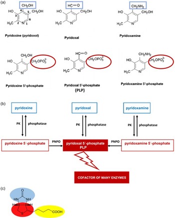
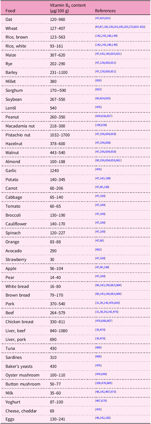
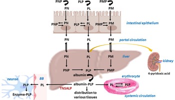
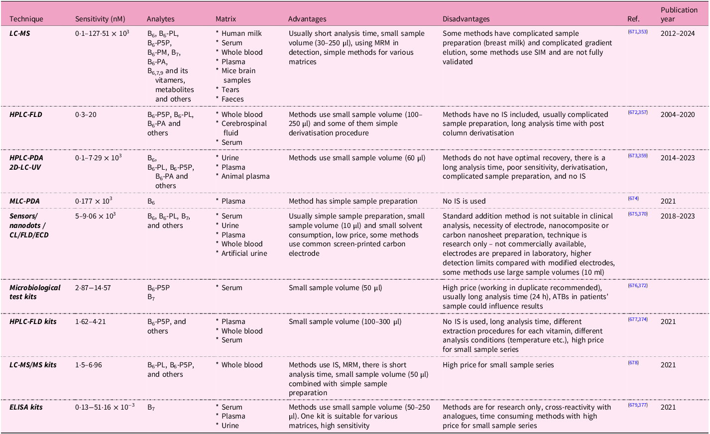
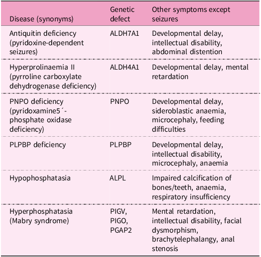
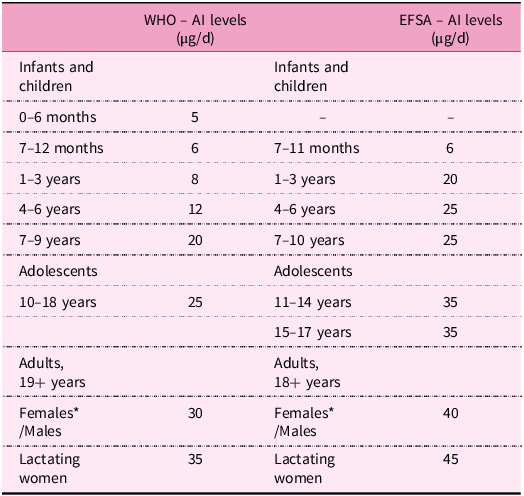
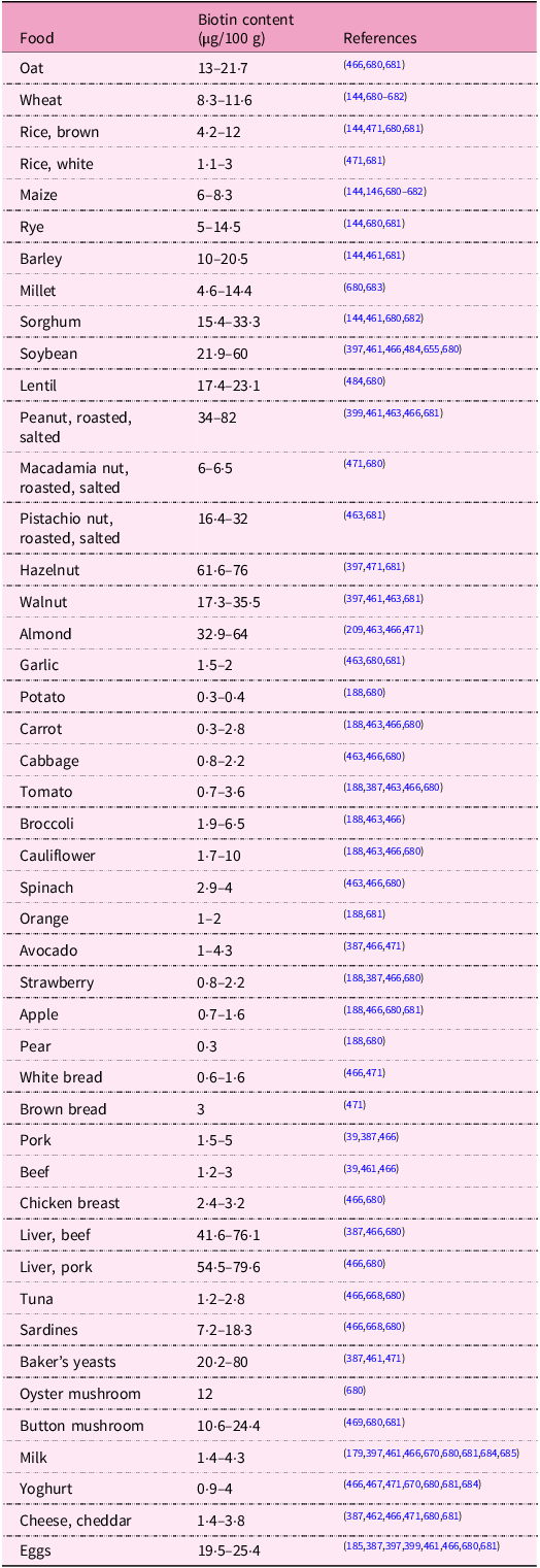
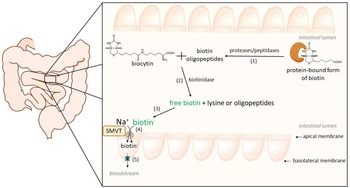
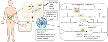
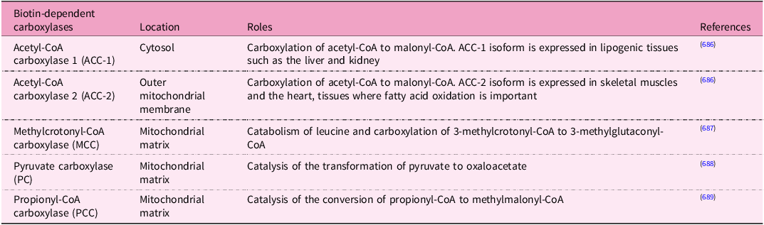
References

Hrubša, M, Siatka, T, Nejmanová, I, et al. (2022) Biological properties of vitamins of the B-complex, part 1: Vitamins B1, B2, B3, and B5. Nutrients 14, 484.CrossRefGoogle Scholar
McCormick, D (2006) Vitamin B6. In Bowman, B & Russell, R, (eds), Present Knowledge in Nutrition. 9 ed. Washington, DC: International Life Sciences Institute, pp 269277.Google Scholar
Mooney, S, Leuendorf, J-E, Hendrickson, C & Hellmann, H (2009) Vitamin B6: a long known compound of surprising complexity. Molecules 14, 329351.CrossRefGoogle ScholarPubMed
Eliot, A & Kirsch, J (2004) Pyridoxal phosphate enzymes: mechanistic, structural, and evolutionary considerations. Annu Rev Biochem 73, 383415.CrossRefGoogle ScholarPubMed
Leklem, JE (1990) Vitamin B-6: a status report. J Nutr 120 (suppl_11), 15031507.CrossRefGoogle ScholarPubMed
Tanaka, T, Tateno, Y & Gojobori, T (2005) Evolution of vitamin B6 (pyridoxine) metabolism by gain and loss of genes. Mol Biol Evol 22, 243250.CrossRefGoogle ScholarPubMed
Hellmann, H & Mooney, S (2010) Vitamin B6: a molecule for human health? Molecules 15, 442459.CrossRefGoogle ScholarPubMed
Parra, M, Stahl, S & Hellmann, H (2018) Vitamin B6 and its role in cell metabolism and physiology. Cells 7, 84.CrossRefGoogle ScholarPubMed
Tambasco-Studart, M, Titiz, O, Raschle, T, Forster, G, Amrhein, N & Fitzpatrick, TB (2005) Vitamin B6 biosynthesis in higher plants. Proc Natl Acad Sci 102, 1368713692.CrossRefGoogle ScholarPubMed
Titiz, O, Tambasco-Studart, M, Warzych, E, et al. (2006) PDX1 is essential for vitamin B6 biosynthesis, development and stress tolerance in Arabidopsis. Plant J 48, 933946.CrossRefGoogle ScholarPubMed
Mooney, S & Hellmann, H (2010) Vitamin B6: killing two birds with one stone? Phytochemistry 71, 495501.CrossRefGoogle ScholarPubMed
Fitzpatrick, TB (2011) Vitamin B6 in plants: more than meets the eye. Adv Bot Res 59, 138.CrossRefGoogle Scholar
Fitzpatrick, TB, Amrhein, N, Kappes, B, Macheroux, P, Tews, I & Raschle, T (2007) Two independent routes of de novo vitamin B6 biosynthesis: not that different after all. Biochem J 407, 113.CrossRefGoogle Scholar
Roje, S (2007) Vitamin B biosynthesis in plants. Phytochemistry 68, 19041921.CrossRefGoogle ScholarPubMed
Gerdes, S, Lerma-Ortiz, C, Frelin, O, et al. (2012) Plant B vitamin pathways and their compartmentation: a guide for the perplexed. J Exp Bot 63, 53795395.CrossRefGoogle Scholar
Leuendorf, JE, Osorio, S, Szewczyk, A, Fernie, AR & Hellmann, H (2010) Complex assembly and metabolic profiling of Arabidopsis thaliana plants overexpressing vitamin B6 biosynthesis proteins. Mol Plant 3, 890903.CrossRefGoogle ScholarPubMed
Mittenhuber, G (2001) Phylogenetic analyses and comparative genomics of vitamin B6 (pyridoxine) and pyridoxal phosphate biosynthesis pathways. J Mol Microbiol Biotechnol 3, 120.Google ScholarPubMed
Kondo, H, Nakamura, Y, Dong, Y-X, Nikawa, J-I & Sueda, S (2004) Pyridoxine biosynthesis in yeast: participation of ribose 5-phosphate ketol-isomerase. Biochem J 379, 6570.CrossRefGoogle ScholarPubMed
Sakai, A, Kita, M & Tani, Y (2004) Recent progress of vitamin B6 biosynthesis. J Nutr Sci Vitaminol 50, 6977.CrossRefGoogle ScholarPubMed
Tramonti, A, Nardella, C, di Salvo, ML, et al. (2021) Knowns and unknowns of vitamin B6 metabolism in Escherichia coli. EcoSal Plus 9, eESP-0004.CrossRefGoogle ScholarPubMed
Begley, TP, Chatterjee, A, Hanes, JW, Hazra, A & Ealick, SE (2008) Cofactor biosynthesis—still yielding fascinating new biological chemistry. Curr Opin Chem Biol 12, 118125.CrossRefGoogle ScholarPubMed
Magnusdottir, S, Ravcheev, D, de Crecy-Lagard, V & Thiele, I (2015) Systematic genome assessment of B-vitamin biosynthesis suggests co-operation among gut microbes. Front Genet 6, 148.CrossRefGoogle ScholarPubMed
Liu, Z, Farkas, P, Wang, K, Kohli, MO & Fitzpatrick, TB (2022) B vitamin supply in plants and humans: the importance of vitamer homeostasis. Pl J 111, 662682.CrossRefGoogle ScholarPubMed
Stach, K, Stach, W & Augoff, K (2021) Vitamin B6 in health and disease. Nutrients 13, 3229.CrossRefGoogle ScholarPubMed
Meir, Z & Osherov, N (2018) Vitamin biosynthesis as an antifungal target. J Fungi 4, 72.CrossRefGoogle Scholar
Scott, DE, Ciulli, A & Abell, C (2007) Coenzyme biosynthesis: enzyme mechanism, structure and inhibition. Nat Prod Rep 24, 10091026.CrossRefGoogle ScholarPubMed
Chen, H & Xiong, L (2009) Enhancement of vitamin B6 levels in seeds through metabolic engineering. Plant Biotechnol J 7, 673681.CrossRefGoogle ScholarPubMed
da Silva, VR & Gregory, JF (2020) Chapter 13 – vitamin B6. In Marriott, BP, Birt, DF, Stallings, VA & Yates, AA, (eds), Present knowledge in nutrition, 11th ed. London, UKAcademic Press, pp 225237.CrossRefGoogle Scholar
EFSA (2016) Dietary reference values for vitamin B6. EFSA J14, e04485.Google Scholar
Chawla, J and Kvarnberg, D (2014) Hydrosoluble vitamins. In Biller, J & Ferro, JM (eds), Handbook of clinical neurology, neurologic aspects of systemic disease part II, 1st ed. Amsterdam, Netherlands: Elsevier. 120, pp 891914.Google Scholar
Wyness, L, Weichselbaum, E, O’connor, A, et al. (2011) Red meat in the diet: an update. Nutr Bull 36, 3477.CrossRefGoogle Scholar
Pereira, PMdCC & Vicente, AFdRB (2013) Meat nutritional composition and nutritive role in the human diet. Meat Sci 93, 586592.CrossRefGoogle ScholarPubMed
Iglesia, I, Mouratidou, T, Gonzalez-Gross, M, et al. (2017) Foods contributing to vitamin B6, folate, and vitamin B 12 intakes and biomarkers status in European adolescents: the HELENA study. Eur J Nutr 56, 17671782.CrossRefGoogle ScholarPubMed
Kraemer, K, Semba, RD, Eggersdorfer, M & Schaumberg, DA (2012) Introduction: the diverse and essential biological functions of vitamins. Ann Nutr Metab 61, 185191.CrossRefGoogle ScholarPubMed
Kennedy, DO (2016) B vitamins and the brain: mechanisms, dose and efficacy—a review. Nutrients 8, 68.CrossRefGoogle ScholarPubMed
Geiker, NRW, Bertram, HC, Mejborn, H, et al. (2021) Meat and human health—current knowledge and research gaps. Foods 10, 1556.CrossRefGoogle ScholarPubMed
McDowell, LR (2000) Vitamin B6. In LR McDowell (author), Vitamins in animal and human nutrition, 2nd ed. Ames, USA: Iowa state university Press, pp 385417.*CrossRefGoogle Scholar
Li, C (2017) The role of beef in human nutrition and health. In Dikeman, ME (ed), Ensuring safety and quality in the production of beef. 1st ed., Vol 2. Cambridge, UK: Burleigh Dodds Science Publishing, pp 329338.CrossRefGoogle Scholar
Ahmad, RS, Imran, A & Hussain, MB (2018) Nutritional composition of meat. In Arshad, MS (ed), Meat science and nutrition. London, UK: IntechOpen Limited. https://www.intechopen.com/chapters/61245 Available from: http://dx.doi.org/10.5772/intechopen.77045.Google Scholar
Hall, APB, Moore, JG & Morgan, AF (1956) Vitamin content of avocados. Calif Agric 10, 1314.Google Scholar
Mielgo-Ayuso, J, Aparicio-Ugarriza, R, Olza, J, et al. (2018) Dietary intake and food sources of niacin, riboflavin, thiamin and vitamin B6 in a representative sample of the Spanish population. The ANIBES study. Nutrients 10, 846.CrossRefGoogle Scholar
Neufingerl, N & Eilander, A (2020) Nutrient intake and status in adults consuming plant-based diets compared to meat-eaters: a systematic review. Nutrients 14, 29.CrossRefGoogle Scholar
Yoshino, K, Inagawa, M, Oshima, M, et al. (2005) Trends in dietary intake of folate, vitamins B6, and B12 among Japanese adults in two rural communities from 1974 through 2001. J Epidemiol 15, 2937.CrossRefGoogle ScholarPubMed
Jungert, A, Linseisen, J, Wagner, K-H, Richter, M & German Nutrition Society (2020) Revised DA-CH reference values for the intake of vitamin B6. Ann Nutr Metab 76, 213222.CrossRefGoogle Scholar
Shewry, PR & Hey, SJ (2015) The contribution of wheat to human diet and health. Food Energy Secur 4, 178202.CrossRefGoogle ScholarPubMed
Waldmann, A, Dörr, B, Koschizke, J, Leitzmann, C & Hahn, A (2006) Dietary intake of vitamin B6 and concentration of vitamin B6 in blood samples of German vegans. Public Health Nutr 9, 779784.CrossRefGoogle ScholarPubMed
Köseoğlu, SZA (2020) Determination and evaluation of the pyridoxal, pyridoxine, and pyridoxamine forms of vitamin B-6 in plant-based foods in terms of healthy vegetarian nutrition. Prog Nutr 22, e2020015.Google Scholar
Yaman, M (2019) Determination and evaluation in terms of healthy nutrition of the pyridoxal, pyridoxine and pyridoxamine forms of vitamin B6 in animal-derived foods. Eur J Sci Technol, 2019, 611617. https://dergipark.org.tr/tr/download/article-file/683971.CrossRefGoogle Scholar
Melanson, KJ, Angelopoulos, TJ, Nguyen, VT, et al. (2006) Consumption of whole-grain cereals during weight loss: effects on dietary quality, dietary fiber, magnesium, vitamin B-6, and obesity. J Am Diet Assoc 106, 13801388.CrossRefGoogle ScholarPubMed
Planells, E, Sanchez, C, Montellano, M, Mataix, J & Llopis, J (2003) Vitamins B6 and B12 and folate status in an adult Mediterranean population. Eur J Clin Nutr 57, 777785.CrossRefGoogle Scholar
EFSA Panel on Dietetic Products, Nutrition and Allergies (2016) Allergies. Dietary reference values for vitamin B6. EFSA J 14, e04485.Google Scholar
Rybicka, I & Gliszczynska-Swiglo, A (2017) Gluten-free flours from different raw materials as the source of vitamin B1, B2, B3 and B6. J Nutr Sci Vitaminol 63, 125132.CrossRefGoogle Scholar
Kamyab, S, Gharachorloo, M, Honarvar, M & Ghavami, M (2020) Quantitative analysis of bioactive compounds present in Iranian royal jelly. J Apic Res 59, 4252.CrossRefGoogle Scholar
Ollilainen, V-M (1999) HPLC analysis of vitamin B6 in foods. Agric Food Sci 8, 515619.CrossRefGoogle Scholar
Ndaw, S, Bergaentzle, M, Aoude-Werner, D & Hasselmann, C (2000) Extraction procedures for the liquid chromatographic determination of thiamin, riboflavin and vitamin B6 in foodstuffs. Food Chem 71, 129138.CrossRefGoogle Scholar
Schmidt, A, Schreiner, M & Mayer, H (2017) Rapid determination of the various native forms of vitamin B6 and B2 in cow’s milk using ultra-high performance liquid chromatography. J Chromator A 1500, 8995.CrossRefGoogle ScholarPubMed
Reynolds, RD (1988) Bioavailability of vitamin B-6 from plant foods. Am J Clin Nutr 48, 863867.CrossRefGoogle ScholarPubMed
Ink, SL, Gregory, JF III & Sartain, DB (1986) Determination of pyridoxine beta-glucoside bioavailability using intrinsic and extrinsic labeling in the rat. J Agric Food Chem 34, 857862.CrossRefGoogle Scholar
Gregory, J III, Ink, S & Sartain, D (1986) Degradation and binding to food proteins of vitamin B-6 compounds during thermal processing. J Food Sci 51, 13451351.Google Scholar
EFSA (2023) Scientific opinion on the tolerable upper intake level for vitamin B6. EFSA J 21, e08006.Google Scholar
Kabir, H, Leklem, JE & Miller, LT (1983) Comparative vitamin B-6 bioavailability from tuna, whole wheat bread and peanut butter in humans. J Nutr 113, 24122420.CrossRefGoogle ScholarPubMed
Gregory, JF III (1998) Nutritional properties and significance of vitamin glycosides. Annu Rev Nutr 18, 277296.CrossRefGoogle ScholarPubMed
Gregory, JF III & Ink, SL (1987) Identification and quantification of pyridoxine beta-glucoside as a major form of vitamin B6 in plant-derived foods. J Agric Food Chem 35, 7682.CrossRefGoogle Scholar
Gregory, JR III (1997) Bioavailability of vitamin B-6. Eur J Clin Nutr 51, S43S8.Google ScholarPubMed
Kabir, H, Leklem, J & Miller, LT (1983) Measurement of glycosylated vitamin B6 in foods. J Food Sci 48, 14221425.CrossRefGoogle Scholar
Kall, MA (2003) Determination of total vitamin B6 in foods by isocratic HPLC: a comparison with microbiological analysis. Food Chem 82, 315327.CrossRefGoogle Scholar
Bachmann, T, Maurer, A & Rychlik, M (2020) Development of a LC-MS/MS method using stable isotope dilution for the quantification of individual B6 vitamers in fruits, vegetables, and cereals. Anal Bioanal Chem 412, 72377252.CrossRefGoogle ScholarPubMed
Mangel, N, Fudge, JB, Gruissem, W, Fitzpatrick, TB & Vanderschuren, H (2022) Natural variation in vitamin B1 and vitamin B6 contents in rice germplasm. Front Plant Sci 13, 856880.CrossRefGoogle ScholarPubMed
Gregory, JF III, Trumbo, PR, Bailey, LB, Toth, JP, Baumgartner, TG & Cerda, JJ (1991) Bioavailability of pyridoxine-5′-β-d-glucoside determined in humans by stable-isotopic methods. J Nutr 121, 177186.CrossRefGoogle ScholarPubMed
Gregory, J III (2012) Accounting for differences in the bioactivity and bioavailability of vitamers. Food Nutr Res 56, 5809.CrossRefGoogle ScholarPubMed
Bird, RP (2018) The emerging role of vitamin B6 in inflammation and carcinogenesis. Adv Food Nutr Res 83, 151194.CrossRefGoogle ScholarPubMed
Sokač, DG, Kovač, S, Bušić, V, Martin, CR & Furač, JV (2012) The chemistry and biochemistry of vitamin B6: synthesis of novel analogues of vitamin B6. In Preedy VR (ed), B vitamins and folate: chemistry, analysis, function and effects. Cambridge, UK: The Royal Society of Chemistry. pp 135145.CrossRefGoogle Scholar
Nakano, H & McMahon, LG & Gregory, JF III (1997) Pyridoxine-5′-β-d-glucoside exhibits incomplete bioavailability as a source of vitamin B-6 and partially inhibits the utilization of co-ingested pyridoxine in humans. J Nutr 127, 15081513.CrossRefGoogle ScholarPubMed
Armada, LJ, Mackey, AD & Gregory, JF III (2002) Intestinal brush border membrane catalyzes hydrolysis of pyridoxine-5′-β-d-glucoside and exhibits parallel developmental changes of hydrolytic activities toward pyridoxine-5′-β-d-glucoside and lactose in rats. J Nutr 132, 26952699.CrossRefGoogle ScholarPubMed
Roth-Maier, DA, Kettler, SI & Kirchgessner, M (2002) Availability of vitamin B6 from different food sources. Int J Food Sci Nutr 53, 171179.CrossRefGoogle ScholarPubMed
Tsuge, H, Maeno, M, Hayakawa, T & Suzuki, Y (1996) Comparative study of pyridoxine-α, β-glucosides, and phosphopyridoxyl-lysine as a vitamin B6 nutrient. J Nutr Sci Vitaminol 42, 377386.CrossRefGoogle ScholarPubMed
Mackey, AD, Lieu, SO, Carman, C & Gregory, JF III (2003) Hydrolytic activity toward pyridoxine-5′-β-d-glucoside in rat intestinal mucosa is not increased by vitamin B-6 deficiency: effect of basal diet composition and pyridoxine intake. J Nutr 133, 13621367.CrossRefGoogle Scholar
Hansen, CM, Leklem, JE & Miller, LT (1996) Vitamin B-6 status indicators decrease in women consuming a diet high in pyridoxine glucoside. J Nutr 126, 25122518.CrossRefGoogle ScholarPubMed
Schorgg, P, Bärnighausen, T, Rohrmann, S, Cassidy, A, Karavasiloglou, N & Kühn, T (2021) Vitamin B6 status among vegetarians: findings from a population-based survey. Nutrients 13, 1627.CrossRefGoogle ScholarPubMed
Basu, TK & Donaldson, D (2003) Intestinal absorption in health and disease: micronutrients. Best Pract Res Clin Gastroenterol 17, 957979.CrossRefGoogle ScholarPubMed
EFSA (2017) Dietary reference values for nutrients. Summary Report EFSA Supporting Publication 14, p e15121.Google Scholar
Yaman, M, Çatak, J, Uğur, H, et al. (2021) The bioaccessibility of water-soluble vitamins: a review. Trends Food Sci Technol 109, 552563.CrossRefGoogle Scholar
Strain, J, Hughes, C, Pentieva, K, Ward, M, Hoey, L & McNulty, H (2017) The B-vitamins. In Biesalski, HK, Drewnowski, A, Dwyer, JT, Strain, J, Weber, P & Eggersdorfer, M (eds), Sustainable nutrition in a changing world, 1st ed. Cham, Switzerland: Springer International Publishing AG, pp 185205.CrossRefGoogle Scholar
Spinneker, A, Sola, R, Lemmen, V, Castillo, M, Pietrzik, K & Gonzalez-Gross, M (2007) Vitamin B6 status, deficiency and its consequences-an overview. Nutr Hosp 22, 724.Google ScholarPubMed
Yaman, M & Mızrak, ÖF (2019) Determination and evaluation of in vitro bioaccessibility of the pyridoxal, pyridoxine, and pyridoxamine forms of vitamin B6 in cereal-based baby foods. Food Chem 298, 125042.CrossRefGoogle ScholarPubMed
Tarr, JB, Tamura, T & Stokstad, EL (1981) Availability of vitamin B6 and pantothenate in an average American diet in man. Am J Clin Nutr 34, 13281337.CrossRefGoogle Scholar
Combs, G Jr & McClung, J (2017) Chapter 14-vitamin B6. In GF Combs & JP McClung (authors), The vitamins, 5th ed. Fundamental Aspects in Nutrition and Health, London, UK: Academic Press. pp 351370.CrossRefGoogle Scholar
Said, HM (2011) Intestinal absorption of water-soluble vitamins in health and disease. Biochem J 437, 357372.CrossRefGoogle ScholarPubMed
Said, ZM, Subramanian, VS, Vaziri, ND & Said, HM (2008) Pyridoxine uptake by colonocytes: a specific and regulated carrier-mediated process. Am J Physiol-Cell Physiol 294, C1192C1197.CrossRefGoogle ScholarPubMed
Uebanso, T, Shimohata, T, Mawatari, K & Takahashi, A (2020) Functional roles of B-vitamins in the gut and gut microbiome. Mol Nutr Food Res 64, 2000426.CrossRefGoogle ScholarPubMed
Yoshii, K, Hosomi, K, Sawane, K & Kunisawa, J (2019) Metabolism of dietary and microbial vitamin B family in the regulation of host immunity. Front Nutr 6, 48.CrossRefGoogle ScholarPubMed
Rowland, I, Gibson, G, Heinken, A, et al. (2018) Gut microbiota functions: metabolism of nutrients and other food components. Eur J Nutr 57, 124.CrossRefGoogle ScholarPubMed
Rodionov, DA, Arzamasov, AA, Khoroshkin, MS, et al. (2019) Micronutrient requirements and sharing capabilities of the human gut microbiome. Front Microbiol 10, 1316.CrossRefGoogle ScholarPubMed
Mayengbam, S, House, J & Aliani, M (2016) Investigation of vitamin B inadequacy, induced by exposure to the anti-B factor 1-amino D-proline, on plasma lipophilic metabolites of rats: a metabolomics approach. Eur J Nutr 55, 12131223.CrossRefGoogle Scholar
Wada, K, Ishigaki, S, Ueda, K, Sakata, M & Haga, M (1985) An antivitamin B6, 4’-methoxypyridoxine, from the seed of Ginkgo biloba L. Chem Pharm Bull 33, 35553557.CrossRefGoogle ScholarPubMed
Zou, M, Zhang, W, Wu, R, Jiang, H, Cao, F & Su, E (2021) Removal of ginkgotoxin from the Ginkgo biloba seeds powder by adopting membrane separation technology. J Clean Prod 280, 124452.CrossRefGoogle Scholar
Kosaki, Y, Naito, H, Nojima, T & Nakao, A (2020) Epileptic seizure from ginkgo nut intoxication in an adult. Case Rep Emerg Med 2020, ID 5072954.Google ScholarPubMed
Azuma, F, Nokura, K, Kako, T, Kobayashi, D, Yoshimura, T & Wada, K (2020) An adult case of generalized convulsions caused by the ingestion of Ginkgo biloba seeds with alcohol. Intern Med 59, 15551558.CrossRefGoogle ScholarPubMed
Kobayashi, D, Yoshimura, T, Johno, A, Ishikawa, M, Sasaki, K & Wada, K (2015) Decrease in pyridoxal-5′-phosphate concentration and increase in pyridoxal concentration in rat plasma by 4′-O-methylpyridoxine administration. Nutr Res 35, 637642.CrossRefGoogle ScholarPubMed
Kobayashi, D, Yoshimura, T, Johno, A, Sasaki, K & Wada, K (2011) Toxicity of 4′-O-methylpyridoxine-5′-glucoside in Ginkgo biloba seeds. Food Chem 126, 11981202.CrossRefGoogle Scholar
Leistner, E & Drewke, C (2010) Ginkgo biloba and ginkgotoxin. J Nat Prod 73, 8692.CrossRefGoogle ScholarPubMed
Kajiyama, Y, Fujii, K, Takeuchi, H & Manabe, Y (2002) Ginkgo seed poisoning. Pediatrics 109, 325327.CrossRefGoogle ScholarPubMed
Arenz, A, Klein, M, Fiehe, K, et al. (1996) Occurrence of neurotoxic 4′-O-methylpyridoxine in Ginkgo biloba leaves, Ginkgo medications and Japanese Ginkgo food. Planta Med 62, 548551.CrossRefGoogle ScholarPubMed
Buss, K, Drewke, C, Lohmann, S, Piwonska, A & Leistner, E (2001) Properties and interaction of heterologously expressed glutamate decarboxylase isoenzymes GAD65kDa and GAD67kDa from human brain with ginkgotoxin and its 5´-phosphate. J Med Chem 44, 31663174.CrossRefGoogle ScholarPubMed
Kästner, U, Hallmen, C, Wiese, M, Leistner, E & Drewke, C (2007) The human pyridoxal kinase, a plausible target for ginkgotoxin from Ginkgo biloba . FEBS J 274, 10361045.CrossRefGoogle ScholarPubMed
Samuels, N, Finkelstein, Y, Singer, SR & Oberbaum, M (2008) Herbal medicine and epilepsy: proconvulsive effects and interactions with antiepileptic drugs. Epilepsia 49, 373380.CrossRefGoogle ScholarPubMed
Mei, N, Guo, X, Ren, Z, Kobayashi, D, Wada, K & Guo, L (2017) Review of Ginkgo biloba-induced toxicity, from experimental studies to human case reports. J Environ Sci Health, Part C 35, 128.CrossRefGoogle ScholarPubMed
Zhu, J-P, Gong, H, Labreche, F, et al. (2021) In vivo toxicity assessment of 4′-O-methylpyridoxine from Ginkgo biloba seeds: growth, hematology, metabolism, and oxidative parameters. Toxicon 201, 6673.CrossRefGoogle ScholarPubMed
Zhu, J-P, Gong, H, Wu, C-E, Fan, G-J, Li, T-T & Wang, J-H (2021) 4′-O-methylpyridoxine: preparation from Ginkgo biloba seeds and cytotoxicity in GES-1 cells. Toxins (Basel) 13, 95.CrossRefGoogle ScholarPubMed
Zhang, W, Shi, M, Zhang, F, Cao, F & Su, E (2021) A facile method to determine the native contents of 4′-O-methylpyridoxine and 4′-O-methylpyridoxine-5′-glucoside in Ginkgo biloba seeds. J Agric Food Chem 69, 1427014277.CrossRefGoogle ScholarPubMed
Lim, HB & Kim, D (2018) Effect of heat treatment on 4′-O-methylpyridoxine (MPN) content in Ginkgo biloba seed extract solution. J Sci Food Agric 98, 51535156.CrossRefGoogle ScholarPubMed
Gong, H, Wu, C-E, Fan, G-J, Li, T-T, Wang, J-H & Wang, T (2018) Determination and comparison of 4′-O-methylpyridoxine analogues in Ginkgo biloba seeds at different growth stages. J Agric Food Chem 66, 79167922.CrossRefGoogle ScholarPubMed
Steyn, PS, Vleggaar, R & Anderson, LA (1987) Structure elucidation of two neurotoxins from Albizia tanganyicensis. S Afr J Chem 40, 191192.Google Scholar
Mayengbam, S, Raposo, S, Aliani, M & House, JD (2015) Oral exposure to the anti-pyridoxine compound 1-amino d-proline further perturbs homocysteine metabolism through the transsulfuration pathway in moderately vitamin B6 deficient rats. J Nutr Biochem 26, 241249.CrossRefGoogle Scholar
Mayengbam, S, Raposo, S, Aliani, M, House, JD (2016) A vitamin B-6 antagonist from flaxseed perturbs amino acid metabolism in moderately vitamin B-6-deficient male rats. J Nutr 146, 1420.CrossRefGoogle ScholarPubMed
Mayengbam, S, Yang, H, Barthet, V, Aliani, M & House, JD (2014) Identification characterization, and quantification of an anti-pyridoxine factor from flaxseed using ultrahigh-performance liquid chromatography–mass spectrometry. J Agric Food Chem 62, 419426.CrossRefGoogle ScholarPubMed
Sasaoka, K, Ogawa, T, Moritoki, K & Kimoto, M (1976) Antivitamin B-6 effect of 1-aminoproline on rats. Biochim Biophys Acta Gen Sub 428, 396402.CrossRefGoogle ScholarPubMed
Klosterman, HJ (1974) Vitamin B6 antagonists of natural origin. J Agric Food Chem 22, 1316.CrossRefGoogle ScholarPubMed
Erogul, M, Ernsting, K & Savitt, D (2001) Gyromitra mushrooms. In MA McGuigan, A. Anderson & A Woolf A (eds), Clin Toxicol Rev 23. Boston, USA: Massachusetts/Rhode Island Poison Control System.Google Scholar
White, J, Weinstein, SA, De Haro, L, et al. (2019) Mushroom poisoning: a proposed new clinical classification. Toxicon 157, 5365.CrossRefGoogle ScholarPubMed
Lagrange, E & Vernoux, J-P (2020) Warning on false or true morels and button mushrooms with potential toxicity linked to hydrazinic toxins: an update. Toxins (Basel) 12, 482.CrossRefGoogle ScholarPubMed
Horowitz, KM & Kong, EL & Horowitz, BZ (2017) Gyromitra mushroom toxicity. StatPearls Publishing. Available from: https://www.ncbi.nlm.nih.gov/books/NBK430685/.Google Scholar
Berdanier, CD (2007) Food constituents. In Berdanier, CD, Dwyer, JT & Feldman, EB (eds), Handbook of nutrition and food. Boca Raton, USA: CRC Press, pp 335.CrossRefGoogle Scholar
Arshadi, M, Nilsson, C & Magnusson, B (2006) Gas chromatography–mass spectrometry determination of the pentafluorobenzoyl derivative of methylhydrazine in false morel (Gyromitra esculenta) as a monitor for the content of the toxin gyromitrin. J Chromator A 1125, 229233.CrossRefGoogle ScholarPubMed
Roupas, P, Keogh, J, Noakes, M, Margetts, C & Taylor, P (2010) Mushrooms and agaritine: a mini-review. J Funct Foods 2, 9198.CrossRefGoogle Scholar
Klosterman, HJ (1979) Vitamin B6 antagonists of natural origin. Methods Enzymol 62, 483495.CrossRefGoogle ScholarPubMed
Negi, VS, Pal, A & Borthakur, D (2021) Biochemistry of plants N–heterocyclic non-protein amino acids. Amino Acids 53, 801812.CrossRefGoogle ScholarPubMed
Honda, MD & Borthakur, D (2019) Mimosine concentration in Leucaena leucocephala under various environmental conditions. Trop Grass-Forra Trop 7, 164172.CrossRefGoogle Scholar
Oogai, S, Fukuta, M, Inafuku, M & Oku, H (2022) Isolation and characterization of mimosine degrading enzyme from Arthrobacter sp. Ryudai-S1. World J Microbiol Biotechnol 38, 172.CrossRefGoogle ScholarPubMed
Rosenthal, GA (1981) A mechanism of L-canaline toxicity. Eur J Biochem 114, 301304.CrossRefGoogle ScholarPubMed
Rosenthal, GA (1997) L-canaline: a potent antimetabolite and anti-cancer agent from leguminous plants. Life Sci 60, 16351641.CrossRefGoogle ScholarPubMed
Gregory, J III & Kirk, J (1981) The bioavailability of vitamin B6 in foods. Nutr Rev 9, 18.Google Scholar
Özilgen, S & Özilgen, M (2017) General template for the FMEA applications in primary food processing. In Hitzmann, B (ed), Measurement, modeling and automation in advanced food processing. Cham: Springer International Publishing, pp 2969.Google Scholar
Berry Ottaway, P (2010) Stability of vitamins during food processing and storage. In Skibsted, LH, Risbo, J & Andersen, ML (eds), Chemical deterioration and physical instability of food and beverages. Cambridge, UK: Woodhead Publishing, pp 539560.CrossRefGoogle Scholar
Godoy, HT, Amaya-Farfan, J & Rodriguez-Amaya, DB (2021) Degradation of vitamins. In Rodriguez-Amaya, DB & Amaya-Farfan, J (eds), Chemical changes during processing and storage of foodsLondon, UK: Academic Press, pp 329383.CrossRefGoogle Scholar
Slavin, JL, Jacobs, D & Marquart, L (2000) Grain processing and nutrition. Crit Rev Food Sci Nutr 40, 309326.CrossRefGoogle ScholarPubMed
Thielecke, F, Lecerf, J-M & Nugent, AP (2021) Processing in the food chain: do cereals have to be processed to add value to the human diet? Nutr Res Rev 34, 159173.CrossRefGoogle Scholar
Călinoiu, LF & Vodnar, DC (2018) Whole grains and phenolic acids: a review on bioactivity, functionality, health benefits and bioavailability. Nutrients 10, 1615.CrossRefGoogle ScholarPubMed
Batifoulier, F, Verny, M-A, Chanliaud, E, Remesy, C & Demigné, C (2006) Variability of B vitamin concentrations in wheat grain, milling fractions and bread products. Eur J Agron 25, 163169.CrossRefGoogle Scholar
Lebert, L, Buche, F, Sorin, A & Aussenac, T (2022) The wheat aleurone layer: optimisation of its benefits and application to bakery products. Foods 11, 3552.CrossRefGoogle ScholarPubMed
Brouns, F, Hemery, Y, Price, R & Anson, NM (2012) Wheat aleurone: separation, composition, health aspects, and potential food use. Crit Rev Food Sci Nutr 52, 553568.CrossRefGoogle ScholarPubMed
Titcomb, TJ & Tanumihardjo, SA (2019) Global concerns with B vitamin statuses: biofortification, fortification, hidden hunger, interactions, and toxicity. Compr Rev Food Sci Food Saf 18, 19681984.CrossRefGoogle ScholarPubMed
Garg, M, Sharma, A, Vats, S, et al. (2021) Vitamins in cereals: a critical review of content, health effects, processing losses, bioaccessibility, fortification, and biofortification strategies for their improvement. Front Nutr 8, 586815.CrossRefGoogle ScholarPubMed
Hegedüs, M, Pedersen, B & Eggum, BO (1985) The influence of milling on the nutritive value of flour from cereal grains. 7. Vitamins and tryptophan. Plant Foods Hum Nutr 35, 175180.CrossRefGoogle Scholar
Gwirtz, JA & Garcia-Casal, MN (2014) Processing maize flour and corn meal food products. Ann N Y Acad Sci 1312, 6675.CrossRefGoogle ScholarPubMed
Dunn, ML, Jain, V & Klein, BP (2014) Stability of key micronutrients added to fortified maize flours and corn meal. Ann N Y Acad Sci 1312, 1525.CrossRefGoogle ScholarPubMed
Henry, C & Heppell, N (2002) Nutritional losses and gains during processing: future problems and issues. Proc Nutr Soc 61, 145148.CrossRefGoogle ScholarPubMed
Tiozon, RJN, Fernie, AR & Sreenivasulu, N (2021) Meeting human dietary vitamin requirements in the staple rice via strategies of biofortification and post-harvest fortification. Trends Food Sci Technol 109, 6582.CrossRefGoogle Scholar
Kyritsi, A, Tzia, C & Karathanos, VT (2011) Vitamin fortified rice grain using spraying and soaking methods. LWT – Food Sci Technol 44, 312320.CrossRefGoogle Scholar
de Pee, S (2014) Proposing nutrients and nutrient levels for rice fortification. Ann N Y Acad Sci 1324, 5566.CrossRefGoogle ScholarPubMed
Bhar, S, Bose, T, Dutta, A & Mande, SS (2022) A perspective on the benefits of consumption of parboiled rice over brown rice for glycaemic control. Eur J Nutr 61, 615624.CrossRefGoogle Scholar
Schroeder, HA (1971) Losses of vitamins and trace minerals resulting from processing and preservation of foods. Am J Clin Nutr 24, 562573.CrossRefGoogle ScholarPubMed
Aboul-Enein, HY & Loutfy, MA (1984) Pyridoxine hydrochloride. In Analytical profiles of drug substances, 13, In K Florey (ed), Analytical profiles of drug substances, 13. Orlando, USA: Academic Press, pp 447486.CrossRefGoogle Scholar
Schnellbaecher, A, Binder, D, Bellmaine, S & Zimmer, A (2019) Vitamins in cell culture media: stability and stabilization strategies. Biotechnol Bioeng 116, 15371555.CrossRefGoogle ScholarPubMed
Yessaad, M, Bernard, L, Bourdeaux, D, Chennell, P & Sautou, V (2018) Development of a stability indicating method for simultaneous analysis of five water-soluble vitamins by liquid chromatography. Pharm Technol Hosp Pharm 3, 207218.CrossRefGoogle Scholar
Juhász, M, Takahashi, S, Kitahara, Y & Fujii, T (2012) Thermal decomposition of pyridoxine: an evolved gas analysis-ion attachment mass spectrometry study. Rap Comm Mas Spect 26, 759764.CrossRefGoogle ScholarPubMed
Ferguson, TI, Emery, S, Price-Davies, R & Cosslett, AG (2014) A review of stability issues associated with vitamins in parenteral nutrition. e-SPEN J 9, e49e53.CrossRefGoogle Scholar
Bonrath, WPK, Zhang, QM, Pauling, H & Weimann, BJ (2020) Vitamins, 10. Vitamin B6. In Bellussi, G, Bohnet, M, Bus, J, Drauz, K, Greim, H, Jackel, KP, Karst, U, Kleemann, A, Kreysa, G, Laird, T, Meier, W, Ottow, E, Roper, M, Scholtz, J, Sundmacher, K, Ulber, R, Wietelmann, U (eds), Ullmann’s encyclopedia of industrial chemistry, 7th ed. Weinheim, Germany: Wiley-VCH Verlag GmbH & Co. KGaA, pp 116.Google Scholar
Gregory, J III & Kirk, J (1978) Assessment of storage effects on vitamin B6 stability and bioavailability in dehydrated food systems. J Food Sci 43, 18011808.CrossRefGoogle Scholar
Gregory, J III & Leatham, K (1990) Lack of vitamin B6 activity of 6-hydroxypyridoxine. J Food Sci 55, 11431146.Google Scholar
Shibata, K, Yasuhara, Y & Yasuda, K (2001) Effects of cooking methods on the retention of vitamin B6 in foods, and the approximate cooking loss in daily meals. J Home Econ Jpn 52, 11871197.Google Scholar
McCormick, DB (1978) Nature and activity of vitamin B6 bound in heat-processed foods. Nutr Rev 36, 346348.Google Scholar
Lešková, E, Kubíková, J, Kováčiková, E, Košická, M, Porubská, J & Holčíková, K (2006) Vitamin losses: retention during heat treatment and continual changes expressed by mathematical models. J Food Compost Anal 19, 252276.CrossRefGoogle Scholar
Fuliaş, A, Vlase, G, Vlase, T, Oneţiu, D, Doca, N & Ledeţi, I (2014) Thermal degradation of B-group vitamins: B1, B2 and B6 . J Therm Anal Calorim 118, 10331038.CrossRefGoogle Scholar
Riaz, MN, Asif, M & Ali, R (2009) Stability of vitamins during extrusion. Crit Rev Food Sci Nutr 49, 361368.CrossRefGoogle ScholarPubMed
Ribeiro, DO, Pinto, DC, Lima, LMT, Volpato, NM, Cabral, LM & de Sousa, VP (2011) Chemical stability study of vitamins thiamine, riboflavin, pyridoxine and ascorbic acid in parenteral nutrition for neonatal use. Nutr J 10, 19.CrossRefGoogle ScholarPubMed
Mašková, E, Rysova, J, Fiedlerova, V & Holasova, M (1994) Vitamin and mineral retention in meat in various cooking methods. Potravinarske Vedy-UZPI (Czech Republic) 12, 407416.Google Scholar
Bognár, A (1993) Studies on the influence of cooking on the vitamin B6 content of food. In U Schlemmer (ed) Bioavailability ’93: nutritional, chemical and food processing implications of nutrient availability Proceedings, Part 1/2. Karlsruhe: Bundesforschungsanstalt für Ernährung S, pp 346351.Google Scholar
Bognár, A (1998) Comparative study of frying to other cooking techniques influence on the nutritive value. Grasas Aceites 49, 250260.CrossRefGoogle Scholar
Bognár, A (2002) Tables on weight yield of food and retention factors of food constituents for the calculation of nutrient composition of cooked foods (dishes). Karlsruhe, Germany: Bundesforschungsanstalt für Ernährung.Google Scholar
Çatak, J & Çaman, R (2020) Pyridoxal pyridoxamine, and pyridoxine cooking loss: characterizing vitamin B6 profiles of chicken meats before and after cooking. J Food Process Preserv 44, e14798.CrossRefGoogle Scholar
USDA (2007) USDA Table of Nutrient Retention Factors, Release 6: Beltsville, USA. https://www.ars.usda.gov/arsuserfiles/80400530/pdf/retn06.pdf.Google Scholar
Engler, PP & Bowers, JA (1976) B-vitamin retention in meat during storage and preparation. A review. J Am Diet Assoc 69, 253257.CrossRefGoogle ScholarPubMed
Meyer, BH, Mysinger, M & Wodarski, L (1969) Pantothenic acid and vitamin B6 in beef. J Am Diet Assoc 54, 122125.CrossRefGoogle ScholarPubMed
Olds, S, Vanderslice, J & Brochetti, D (1993) Vitamin B6 in raw and fried chicken by HPLC. J Food Sci 58, 505507.CrossRefGoogle Scholar
Pearson, A, Burnside, J, Edwards, H, Glasscock, R, Cunha, T & Novak, A (1951) Vitamin losses in drip obtained upon defrosting frozen meat. Food Res 16, 8587.CrossRefGoogle ScholarPubMed
Pearson, A, West, R & Luecke, R (1959) The vitamin and amino acid content of drip obtained upon defrosting frozen pork. Food Res 24, 515519.CrossRefGoogle Scholar
Çatak, J, Çaman, R & Ceylan, Z (2020) Critical vitamin assessment: pyridoxal, pyridoxamine, and pyridoxine levels for three species of raw and cooked fish samples. J Aquat Food Prod Technol 29, 981989.CrossRefGoogle Scholar
Muehlhoff, E, Bennett, A & McMahon, D (2013) Milk and dairy products in human nutrition. Rome, Italy: FAO, pp 4364.Google Scholar
Ryley, J & Kajda, P (1994) Vitamins in thermal processing. Food Chem 49, 119129.CrossRefGoogle Scholar
Asadullah, K-u-N, Tarar, OM, Ali, SA, Jamil, K & Begum, A (2010) Study to evaluate the impact of heat treatment on water soluble vitamins in milk. J Pak Med Assoc 60, 909912.Google ScholarPubMed
Rolls, BA & Porter, JWG (1973) Some effects of processing and storage on the nutritive value of milk and milk products. Proc Nutr Soc 32, 915.CrossRefGoogle ScholarPubMed
Oamen, E, Hansen, A & Swartzel, K (1989) Effect of ultra-high temperature steam injection processing and aseptic storage on labile water-soluble vitamins in milk. J Dairy Sci 72, 614619.CrossRefGoogle Scholar
Amador-Espejo, G, Gallardo-Chacon, J, Nykänen, H, Juan, B & Trujillo, A (2015) Effect of ultra high-pressure homogenization on hydro-and liposoluble milk vitamins. Food Res Int 77, 4954.CrossRefGoogle Scholar
Roe, M, Church, S, Pinchen, H & Finglas, P (2013) Nutrient analysis of eggs: Analytical report. Norwich: Institute of Food Research.Google Scholar
Öhrvik, V, Carlsen, MH, Källman, A & Martinsen, TA (2015) Improving food composition data by standardizing calculation methods. Copenhagen: Nordisk Ministerråd.Google Scholar
Alajaji, SA & El-Adawy, TA (2006) Nutritional composition of chickpea (Cicer arietinum L.) as affected by microwave cooking and other traditional cooking methods. J Food Compos Anal 19, 806812.CrossRefGoogle Scholar
Roe, M, Church, S, Pinchen, H & Finglas, P (2013) Nutrient analysis of fruit and vegetables; analytical report. Norwich: Institute of Food Research.Google Scholar
Rezaei, M, Alizadeh Sani, M, Amini, M, et al. (2022) Influence of cooking process on the content of water-soluble B vitamins in rice marketed in Iran. Food Sci Nutr 10, 460469.CrossRefGoogle ScholarPubMed
Batifoulier, F, Verny, M-A, Chanliaud, E, Rémésy, C & Demigné, C (2022) Effect of different breadmaking methods on thiamine, riboflavin and pyridoxine contents of wheat bread. J Cer Sci 42, 101108.CrossRefGoogle Scholar
Nurit, E, Lyan, B, Pujos-Guillot, E, Branlard, G & Piquet, A (2016) Change in B and E vitamin and lutein, β-sitosterol contents in industrial milling fractions and during toasted bread production. J Cer Sci 69, 290296.CrossRefGoogle Scholar
Mihhalevski, A, Nisamedtinov, I, Hälvin, K, Ošeka, A & Paalme, T (2013) Stability of B-complex vitamins and dietary fiber during rye sourdough bread production. J Cer Sci 57, 3038.CrossRefGoogle Scholar
Athar, N, Hardacre, A, Taylor, G, Clark, S, Harding, R & McLaughlin, J (2006) Vitamin retention in extruded food products. J Food Compost Anal 19, 379383.CrossRefGoogle Scholar
Ekıncı, R (2005) The effect of fermentation and drying on the water-soluble vitamin content of tarhana, a traditional Turkish cereal food. Food Chem 90, 127132.CrossRefGoogle Scholar
Bulló, M, Juanola-Falgarona, M, Hernández-Alonso, P & Salas-Salvadó, J (2015) Nutrition attributes and health effects of pistachio nuts. Br J Nutr 113, S79S93.CrossRefGoogle ScholarPubMed
Stuetz, W, Schlörmann, W & Glei, M (2017) B-vitamins, carotenoids and α-/γ-tocopherol in raw and roasted nuts. Food Chem 221, 222227.CrossRefGoogle ScholarPubMed
Romanini, E, Rastelli, S, Donadini, G, Lambri, M & Bertuzzi, T (2021) Pyridoxine and folates during small and large scale brewing. J Inst Brew 127, 135139.CrossRefGoogle Scholar
Rossi, F, Spigno, G, Luzzani, G, et al. (2021) Effects of the intake of craft or industrial beer on serum homocysteine. Int J Food Sci Nutr 72, 9398.CrossRefGoogle ScholarPubMed
Bertuzzi, T, Mulazzi, A, Rastelli, S, Donadini, G, Rossi, F & Spigno, G (2020) Targeted healthy compounds in small and large-scale brewed beers. Food Chem 310, 125935.CrossRefGoogle ScholarPubMed
Zhang, H, De Silva, D, Dissanayaka, D, Warkentin, TD & Vandenberg, A (2021) Validated B vitamin quantification from lentils by selected reaction monitoring mass spectrometry. Food Chem 359, 129810.CrossRefGoogle ScholarPubMed
Moongngarm, A & Saetung, N (2010) Comparison of chemical compositions and bioactive compounds of germinated rough rice and brown rice. Food Chem 122, 782788.CrossRefGoogle Scholar
Khalil, A & Mansour, E (1995) The effect of cooking, autoclaving and germination on the nutritional quality of faba beans. Food Chem 54, 177182.CrossRefGoogle Scholar
Žilić, S, Basić, Z, Hadži-Tašković Šukalović, V, Maksimović, V, Janković, M & Filipović, M (2014) Can the sprouting process applied to wheat improve the contents of vitamins and phenolic compounds and antioxidant capacity of the flour? Int J Food Sci Technol 49, 10401047.CrossRefGoogle Scholar
Pinheiro, SS, Anunciação, PC, de Morais Cardoso, L, et al. (2021) Stability of B vitamins, vitamin E, xanthophylls and flavonoids during germination and maceration of sorghum (Sorghum bicolor L.). Food Chem 345, 128775.CrossRefGoogle ScholarPubMed
Martin-Belloso, O & Llanos-Barriobero, E (2001) Proximate composition, minerals and vitamins in selected canned vegetables. Eur Food Res Technol 212, 182187.CrossRefGoogle Scholar
Kilcast, D (1994) Effect of irradiation on vitamins. Food Chem 49, 157164.CrossRefGoogle Scholar
Woodside, J (2015) Nutritional aspects of irradiated food. Stewart Postharvest Rev 11, 16.CrossRefGoogle Scholar
Bernaś, E & Jaworska, G. (2016) Vitamins profile as an indicator of the quality of frozen Agaricus bisporus mushrooms. J Food Compos Anal 49, 18.CrossRefGoogle Scholar
Zwart, S, Kloeris, V, Perchonok, M, Braby, L & Smith, S (2009) Assessment of nutrient stability in foods from the space food system after long-duration spaceflight on the ISS. J Food Sci 74, H209H217.CrossRefGoogle ScholarPubMed
Coad, R & Bui, L (2020) Stability of vitamins B1, B2, B6 and E in a fortified military freeze-dried meal during extended storage. Foods 9, 39.CrossRefGoogle Scholar
Williams, PG (1996) Vitamin retention in cook/chill and cook/hot-hold hospital foodservices. J Am Diet Assoc 96, 490498.CrossRefGoogle Scholar
Fischesser, J, Fritsch, H, Gum, AG, Karge, R, Keuper, R (2009) Manufacture of vitamin B6. Patent No. US 7.495,101 B2.Google Scholar
Bachmann, T & Rychlik, M (2018) Synthesis of [13C3]-B6 vitamers labelled at three consecutive positions starting from [13C3]-propionic acid. Molecules 23, 2117.CrossRefGoogle ScholarPubMed
European Commission (2006) Commission Directive 2006/141/EC of 22 December 2006 on infant formulae and follow-on formulae and amending Directive 1999/21/EC. Off J Eur Union 49, 133.Google Scholar
EFSA (2021) Assessment of a feed additive consisting of vitamin B6 (pyridoxine hydrochloride) for all animal species for the renewal of its authorisation (Kaesler Nutrition GmbH). EFSA J 19, e06612.Google Scholar
Bailey, RL, Gahche, JJ, Lentino, CV, et al. (2011) Dietary supplement use in the United States, 2003–2006. J Nutr 141, 261266.CrossRefGoogle ScholarPubMed
Berendsen, AA, van Lieshout, LE, van den Heuvel, EG, Matthys, C, Péter, S & de Groot, LC (2016) Conventional foods, followed by dietary supplements and fortified foods, are the key sources of vitamin D, vitamin B6, and selenium intake in Dutch participants of the NU-AGE study. Nutr Res 36, 11711181.CrossRefGoogle ScholarPubMed
Bird, JK, Barron, R, Pigat, S & Bruins, MJ (2022) Contribution of base diet, voluntary fortified foods and supplements to micronutrient intakes in the UK. J Nutr Sci 11, e51.CrossRefGoogle ScholarPubMed
Gum, AG (2006) Manufacture of vitamin B6. Patent No. WO 2006/066806 A1.Google Scholar
Eggersdorfer, M, Laudert, D, Letinois, U, et al. (2012) One hundred years of vitamins—a success story of the natural sciences. Angew Chem Int Ed 51, 1296012990.CrossRefGoogle ScholarPubMed
Bonrath, W & Netscher, T (2005) Catalytic processes in vitamins synthesis and production. Appl Catal A: Gen 280, 5573.CrossRefGoogle Scholar
Zou, Y, Shi, X, Zhang, G, Li, Z, Jin, C & Su, W (2013) Improved “oxazole” method for the practical and efficient preparation of pyridoxine hydrochloride (vitamin B6). Org Process Res Dev 17, 14981502.CrossRefGoogle Scholar
Shtyrlin, YG, Petukhov, A, Strelnik, A, et al. (2019) Chemistry of pyridoxine in drug design. Russ Chem Bull 68, 911945.CrossRefGoogle Scholar
Dumond, YR & Gum, AG (2003) Silane reduction of 5-hydroxy-6-methyl-pyridine-3, 4-dicarboxylic acid diethyl ester: synthesis of vitamin B6. Molecules 8, 873881.CrossRefGoogle Scholar
Ledesma-Amaro, RS, Jiménez, A & Revuelta, JL (2013) Microbial production of vitamins. In McNeil, B, Archer, D, Giavasis, I & Harvey, L (eds), Microbial production of food ingredients, enzymes and nutraceuticals. Sawston: Woodhead Publishing, pp 571594.CrossRefGoogle Scholar
Acevedo-Rocha, CG, Gronenberg, LS, Mack, M, Commichau, FM & Genee, HJ (2019) Microbial cell factories for the sustainable manufacturing of B vitamins. Curr Opin Biotechnol 56, 1829.CrossRefGoogle ScholarPubMed
Wang, Y, Liu, L, Jin, Z & Zhang, D (2021) Microbial cell factories for green production of vitamins. Front Bioeng Biotechnol 9, 661562.CrossRefGoogle ScholarPubMed
Commichau, FM, Alzinger, A, Sande, R, et al. (2014) Overexpression of a non-native deoxyxylulose-dependent vitamin B6 pathway in Bacillus subtilis for the production of pyridoxine. Metab Eng 25, 3849.CrossRefGoogle ScholarPubMed
Commichau, FM, Alzinger, A, Sande, R, et al. (2015) Engineering Bacillus subtilis for the conversion of the antimetabolite 4-hydroxy-L-threonine to pyridoxine. Metab Eng 29, 196207.CrossRefGoogle ScholarPubMed
Rosenberg, J, Ischebeck, T & Commichau, FM (2017) Vitamin B6 metabolism in microbes and approaches for fermentative production. Biotechnol Adv 35, 3140.CrossRefGoogle ScholarPubMed
Rosenberg, J, Yeak, KC & Commichau, FM (2018) A two-step evolutionary process establishes a non-native vitamin B6 pathway in Bacillus subtilis . Environ Microbiol 20, 156168.CrossRefGoogle ScholarPubMed
Yamamura, E-T (2019) Bioconversion of pyridoxine to pyridoxamine through pyridoxal using a Rhodococcus expression system. J Biosci Bioeng 127, 7984.CrossRefGoogle ScholarPubMed
Hoshino, T, Ichikawa, K, Nagahashi, Y & Tazoe, M (2004) Microorganism and process for preparing vitamin B6. Patent No. WO 2004/029228 A1.Google Scholar
Ichikawa, K, Hoshino, T & Tazoe, M (1997) Fermentative production of vitamin B6. Patent No. EP 0 765 938 A2.Google Scholar
Tazoe, M, Ichikawa, K & Hoshino, T (1999) Production of vitamin B6 in Rhizobium. Biosci Biotechnol Biochem 63, 13781382.CrossRefGoogle ScholarPubMed
Kim, J, Kershner, JP, Novikov, Y, Shoemaker, RK & Copley, SD (2010) Three serendipitous pathways in E. coli can bypass a block in pyridoxal-5′-phosphate synthesis. Mol Syst Biol 6, 436.CrossRefGoogle Scholar
Richts, B & Commichau, FM (2021) Underground metabolism facilitates the evolution of novel pathways for vitamin B6 biosynthesis. Appl Microbiol Biotechnol 105, 22972305.CrossRefGoogle ScholarPubMed
Cardoso, RV, Fernandes, Â, Gonzaléz-Paramás, AM, Barros, L & Ferreira, IC (2019) Flour fortification for nutritional and health improvement: a review. Food Res Int 125, 108576.CrossRefGoogle ScholarPubMed
Malik, KA & Maqbool, A (2020) Transgenic crops for biofortification. Front Sustain Food Syst 4, 571402.CrossRefGoogle Scholar
Allen, L, De Benoist, B, Dary, O & Hurrell, R (2006) Guidelines on food fortification with micronutrients: WHO/FAO, Geneva, Switzerland: WHO Press.Google Scholar
Bird, JK, Murphy, RA, Ciappio, ED & McBurney, MI (2017) Risk of deficiency in multiple concurrent micronutrients in children and adults in the United States. Nutrients 9, 655.CrossRefGoogle ScholarPubMed
Das, JK, Salam, RA, Mahmood, SB, et al. (2019) Food fortification with multiple micronutrients: impact on health outcomes in general population. Cochrane Database Syst Rev, 2019, CD011400. https://www.cochranelibrary.com/cdsr/doi/10.1002/14651858.CD011400.pub2/epdf/full.Google Scholar
Tucker, KL, Olson, B, Bakun, P, Dallal, GE, Selhub, J & Rosenberg, IH (2004) Breakfast cereal fortified with folic acid, vitamin B-6, and vitamin B-12 increases vitamin concentrations and reduces homocysteine concentrations: a randomized trial. Am J Clin Nutr 79, 805811.CrossRefGoogle ScholarPubMed
Duvenage, SS & Schönfeldt, HC (2007) Impact of South African fortification legislation on product formulation for low-income households. J Food Compos Anal 20, 688695.CrossRefGoogle Scholar
Tang, Y, Xu, M & Shiu-Ming, K (2018) Factors contributing to the high prevalence of vitamin B6 deficiency in US: a systematic review. J Hum Nutr 2, 5864.Google Scholar
Ho, C-L, Quay, TA, Devlin, AM & Lamers, Y (2016) Prevalence and predictors of low vitamin B6 status in healthy young adult women in metro Vancouver. Nutrients 8, 538.CrossRefGoogle ScholarPubMed
Newman, JC, Malek, AM, Hunt, KJ & Marriott, BP (2019) Nutrients in the US diet: naturally occurring or enriched/fortified food and beverage sources, plus dietary supplements: NHANES 2009–2012. J Nutr 149, 14041412.CrossRefGoogle ScholarPubMed
Kim, Y-N & Cho, Y-O (2014) Evaluation of vitamin B6 intake and status of 20- to 64-year-old Koreans. Nutr Res Pract 8, 688694.CrossRefGoogle ScholarPubMed
Pfeiffer, CM, Sternberg, MR, Schleicher, RL, Haynes, BMH, Rybak, ME & Pirkle, JL (2013) The CDC’s second national report on biochemical indicators of diet and nutrition in the u.s. population is a valuable tool for researchers and policy makers. J Nutr 143, 938S947S.CrossRefGoogle ScholarPubMed
Brown, MJ, Ameer, AM, Daley, SF, et al. (2023) Vitamin B6 deficiency. StatPearls [Internet]. Treasure Island, FL: StatPearls Publishing. https://www.ncbi.nlm.nih.gov/books/NBK470579/.Google ScholarPubMed
Morris, MS, Picciano, MF, Jacques, PF & Selhub, J (2008) Plasma pyridoxal 5′-phosphate in the US population: the National Health and Nutrition Examination Survey, 2003–2004 12. Am J Clin Nutr 87, 14461454.CrossRefGoogle Scholar
Bates, CJ, Pentieva, KD, Prentice, A, Mansoor, MA & Finch, S (1999) Plasma pyridoxal phosphate and pyridoxic acid and their relationship to plasma homocysteine in a representative sample of British men and women aged 65 years and over. Br J Nutr 81, 191201.CrossRefGoogle Scholar
Porter, K, Hoey, L, Hughes, CF, Ward, M & McNulty, H (2016) Causes consequences and public health implications of low B-vitamin status in ageing. Nutrients 8, 725.CrossRefGoogle Scholar
ter Borg, S, Verlaan, S, Hemsworth, J, et al. (2015) Micronutrient intakes and potential inadequacies of community-dwelling older adults: a systematic review. Br J Nutr 113, 11951206.CrossRefGoogle ScholarPubMed
FFI. Food Fortification Initiative (2022) https://www.ffinetwork.org/country-profiles.Google Scholar
GFDx. Global Fortification Data Exchange (2022) https://fortificationdata.org/map-number-of-nutrients/.Google Scholar
Swanepoel, E, Havemann-Nel, L, Rothman, M, et al. (2019) Contribution of commercial infant products and fortified staple foods to nutrient intake at ages 6, 12, and 18 months in a cohort of children from a low socio-economic community in South Africa. Matern Child Nutr 15, e12674.CrossRefGoogle Scholar
EU Parliament (2006) Regulation (EC) No 1925/2006 of the European Parliament and of the Council of 20 December 2006 on the addition of vitamins and minerals and of certain other substances to foods. Off J Eur Union 50, 2638.Google Scholar
European Commission (2006) Commission directive 2006/125/EC on processed cereal-based foods and baby foods for infants and young children. Off J Eur Union L339/16.Google Scholar
FSSAI (Food Safety and Standards Authority of India) (2018) Food Safety and Standards (Fortification of Foods) Regulations. In: The Gazette of India: Extraordinarry (PART 111, Section 4). New Delhi, India: Ministry of Health and Family Welfare (MoHFW), Government of India.Google Scholar
Tapola, N, Karvonen, H, Niskanen, L & Sarkkinen, E (2004) Mineral water fortified with folic acid, vitamins B6, B12, D and calcium improves folate status and decreases plasma homocysteine concentration in men and women. Eur J Clin Nutr 58, 376385.CrossRefGoogle ScholarPubMed
Hennessy, A, Walsh, E, Walton, J & Flynn, A (2011) The contribution of fortified foods to micronutrient intake in Irish adults aged 18–64 years. Proc Nutr Soc 70, E112.CrossRefGoogle Scholar
Sharma, S & Litonjua, A (2014) Asthma, allergy, and responses to methyl donor supplements and nutrients. J Allergy Clin Immunol 133, 12461254.CrossRefGoogle ScholarPubMed
Czeizel, AE & Merhala, Z (1998) Bread fortification with folic acid, vitamin B12, and vitamin B6 in hungary. Lancet 352, 1225.CrossRefGoogle ScholarPubMed
Hannon, EM, Kiely, M & Flynn, A (2018) The impact of voluntary fortification of foods on micronutrient intakes in Irish adults. Br J Nutr 97, 11771186.CrossRefGoogle Scholar
Bouis, H (2018) Reducing mineral and vitamin deficiencies through biofortification: progress under HarvestPlus. Hidden hunger: strategies to improve nutrition quality. World Rev Nutr Diet 118, 112122.CrossRefGoogle Scholar
Bouis, HE & Saltzman, A (2017) Improving nutrition through biofortification: a review of evidence from HarvestPlus, 2003 through 2016. Glob Food Sec 12, 4958.CrossRefGoogle ScholarPubMed
Strobbe, S & Van Der Straeten, D (2018) Toward eradication of B-vitamin deficiencies: considerations for crop biofortification. Front Plant Sci 9, 443.CrossRefGoogle ScholarPubMed
Vanderschuren, H, Boycheva, S, Li, K-T, Szydlowski, N, Gruissem, W & Fitzpatrick, TB (2013) Strategies for vitamin B6 biofortification of plants: a dual role as a micronutrient and a stress protectant. Front Plant Sci 4, 143.CrossRefGoogle Scholar
Fudge, J, Mangel, N, Gruissem, W, Vanderschuren, H & Fitzpatrick, TB (2017) Rationalising vitamin B6 biofortification in crop plants. Curr Opin Biotechnol 44, 130137.CrossRefGoogle ScholarPubMed
Blancquaert, D, De Steur, H, Gellynck, X & Van Der Straeten, D (2017) Metabolic engineering of micronutrients in crop plants. Ann N Y Aca Sci 1390, 5973.CrossRefGoogle ScholarPubMed
Shewry, PR, Van Schaik, F, Ravel, C, et al. (2011) Genotype and environment effects on the contents of vitamins B1, B2, B3, and B6 in wheat grain. J Agric Food Chem 59, 1056410571.CrossRefGoogle ScholarPubMed
Mooney, S, Chen, L, Kühn, C, Navarre, R, Knowles, NR & Hellmann, H (2013) Genotype-specific changes in vitamin B6 content and the PDX family in potato. BioMed Res Int 2013, 389723.CrossRefGoogle ScholarPubMed
Azam, FMS, Lian, T, Liang, Q, Wang, W, Zhang, C & Jiang, L (2022) Variation of vitamin B contents in maize inbred lines: potential genetic resources for biofortification. Front Nutr 9, 1029119.CrossRefGoogle Scholar
Herrero, S & Daub, ME (2007) Genetic manipulation of vitamin B-6 biosynthesis in tobacco and fungi uncovers limitations to up-regulation of the pathway. Plant Sci 172, 609620.CrossRefGoogle Scholar
Raschke, M, Boycheva, S, Crèvecoeur, M, et al. (2011) Enhanced levels of vitamin B6 increase aerial organ size and positively affect stress tolerance in Arabidopsis . Plant J 66, 414432.CrossRefGoogle ScholarPubMed
Mohsin, S, Irfan, M, Saeed, A, Malik, KA & Maqbool, A (2022) Enhanced expression of PDX1 accumulates vitamin B6 in transgenic wheat seeds. J Cer Sci 107, 103502.CrossRefGoogle Scholar
Mangel, N, Fudge, JB, Li, KT, et al. (2019) Enhancement of vitamin B6 levels in rice expressing Arabidopsis vitamin B6 biosynthesis de novo genes. Plant J 99, 10471065.CrossRefGoogle ScholarPubMed
Li, K-T, Moulin, M, Mangel, N, et al. (2015) Increased bioavailable vitamin B6 in field-grown transgenic cassava for dietary sufficiency. Nat Biotechnol 33, 10291032.Google ScholarPubMed
Bagri, DS, Upadhyaya, DC, Kumar, A & Upadhyaya, CP (2018) Overexpression of PDX-II gene in potato (Solanum tuberosum L.) leads to the enhanced accumulation of vitamin B6 in tuber tissues and tolerance to abiotic stresses. Plant Sci 272, 267275.CrossRefGoogle Scholar
Coburn, SP, Lewis, DL, Fink, WJ, Mahuren, JD, Schaltenbrand, WE & Costill, DL (1988) Human vitamin B-6 pools estimated through muscle biopsies. Am J Clini Nutr 48, 291294.CrossRefGoogle ScholarPubMed
Hamm, MW, Mehansho, H & Henderson, LM (1979) Transport and metabolism of pyridoxamine and pyridoxamine phosphate in the small intestine of the rat. J Nutr 109, 15521559.CrossRefGoogle ScholarPubMed
Said, HM (2004) Recent advances in carrier-mediated intestinal absorption of water-soluble vitamins. Annu Rev Physiol 66, 419446.CrossRefGoogle ScholarPubMed
Yamashiro, T, Yasujima, T, Said, HM & Yuasa, H (2020) pH-dependent pyridoxine transport by SLC19A2 and SLC19A3: implications for absorption in acidic microclimates. J Biol Chem 295, 1699817008.CrossRefGoogle ScholarPubMed
Surtees, R, Mills, P & Clayton, P (2006) Inborn errors affecting vitamin B6 metabolism. Future Neurol 1, 615620.CrossRefGoogle Scholar
EFSA (2016) Panel on dietetic products, nutrition and allergies, NDA. Dietary reference values for vitamin B6. EFSA J 14, 4485.Google Scholar
Anderson, BB, Fulford-Jones, CE, Child, JA, Beard, ME & Bateman, CJ (1971) Conversion of vitamin B6 compounds to active forms in the red blood cell. J Clin Invest 50, 19011909.CrossRefGoogle ScholarPubMed
Bohney, JP, Fonda, ML & Feldhoff, R (1992) Identification of Lys190 as the primary binding site for pyridoxal 5′-phosphate in human serum albumin. FEBS Lett 298, 266268.CrossRefGoogle Scholar
Fonda, ML, Trauss, C & Guempel, UM (1991) The binding of pyridoxal 5′-phosphate to human serum albumin. Arch Biochem Biophys 288, 7986.CrossRefGoogle ScholarPubMed
Ink, SL & Henderson, L (1984) Effect of binding to hemoglobin and albumin on pyridoxal transport and metabolism. J Biol Chem 259, 58335837.CrossRefGoogle ScholarPubMed
Merrill, AH Jr & Henderson, JM (1990) Vitamin B6 metabolism by human liver. Ann N Y Acad Sci 585, 110117.CrossRefGoogle ScholarPubMed
Merrill, AH Jr, Henderson, JM, Wang, E, McDonald, BW & Millikan, WJ (1984) Metabolism of vitamin B-6 by human liver. J Nutr 114, 16641674.CrossRefGoogle ScholarPubMed
Di Salvo, ML, Contestabile, R & Safo, MK (2011) Vitamin B6 salvage enzymes: mechanism, structure and regulation. Biochim Biophys Acta (BBA)-Proteins Proteomics 1814, 15971608.CrossRefGoogle ScholarPubMed
Lumeng, L & Li, T (1975) Characterization of the pyridoxal 5′-phosphate and pyridoxamine 5′-phosphate hydrolase activity in rat liver. Identity with alkaline phosphatase. J Biol Chem 250, 81268131.CrossRefGoogle ScholarPubMed
Surtees, R, Mills, P & Clayton, P (2006) Inborn errors affecting vitamin B6 metabolism. Future Neurol 1, 615620.CrossRefGoogle Scholar
Stanulović, M, Jeremić, V, Leskovac, V & Chaykin, S (1976) New pathway of conversion of pyridoxal to 4-pyridoxic acid. Enzyme 21, 357369.CrossRefGoogle ScholarPubMed
Phillips, RS (2015) Chemistry and diversity of pyridoxal-5′-phosphate dependent enzymes. Biochim Biophys Acta (BBA)-Proteins Proteomics 1854, 11671174.CrossRefGoogle ScholarPubMed
Brunton, L, Chabner, BA, Knollman, B (2011) Goodman and Gilman’s the pharmacological basis of therapeutics, 12 ed. New York: McGraw-Hill.Google Scholar
Aitken, SM, Lodha, PH & Morneau, DJ (2011) The enzymes of the transsulfuration pathways: active-site characterizations. Biochim Biophys Acta (BBA)-Proteins Proteomics 1814, 15111517.CrossRefGoogle ScholarPubMed
Bender, DA (1999) Non-nutritional uses of vitamin B6. Br J Nutr 81, 720.CrossRefGoogle ScholarPubMed
Ereño-Orbea, J, Majtan, T, Oyenarte, I, Kraus, JP & Martínez-Cruz, LA (2014) Structural insight into the molecular mechanism of allosteric activation of human cystathionine β-synthase by S-adenosylmethionine. Proc Natl Acad Sci 111, E3845E3852.CrossRefGoogle ScholarPubMed
Chang, YC, Scott, R & Graves, D. (1987) Function of pyridoxal 5′-phosphate in glycogen phosphorylase: a model study using 6-fluoro-5′-deoxypyridoxal-and 5′-deoxypyridoxal-reconstituted enzymes. Biochemistry 26, 360367.CrossRefGoogle Scholar
Palm, D, Klein, HW, Schinzel, R, Buehner, M & Helmreich, EJ (1990) The role of pyridoxal 5′-phosphate in glycogen phosphorylase catalysis. Biochemistry 29, 10991107.CrossRefGoogle ScholarPubMed
Takagi, M, Fukui, T & Shimomura, S (1982) Catalytic mechanism of glycogen phosphorylase: pyridoxal (5′) diphospho (1)-alpha-d-glucose as a transition-state analogue. Proc Natl Acad Sci 79, 37163719.CrossRefGoogle Scholar
Bourquin, F, Capitani, G & Grütter, MG (2011) PLP-dependent enzymes as entry and exit gates of sphingolipid metabolism. Protein Sci 20, 14921508.CrossRefGoogle ScholarPubMed
Allen, GF, Neergheen, V, Oppenheim, M, et al. (2010) Pyridoxal 5′-phosphate deficiency causes a loss of aromatic L-amino acid decarboxylase in patients and human neuroblastoma cells, implications for aromatic L-amino acid decarboxylase and vitamin B6 deficiency states. J Neurochem 114, 8796.CrossRefGoogle Scholar
Bayoumi, R & Smith, W (1972) Some effects of dietary vitamin B6 deficiency on γ-aminobutyric acid metabolism in developing rat brain. J Neurochem 19, 18831897.CrossRefGoogle Scholar
Dakshinamurti, K, Sharma, S & Geiger, JD (2003) Neuroprotective actions of pyridoxine. Biochim Biophys Acta (BBA)-Proteins Proteomics 1647, 225229.CrossRefGoogle ScholarPubMed
Stephens, M, Havlicek, V & Dakshinamurti, K (1971) Pyridoxine deficiency and development of the central nervous system in the rat. J Neurochem 18, 24072416.CrossRefGoogle ScholarPubMed
Cervenka, I, Agudelo, L & Kynurenines, RJ (2017) Kynurenines: tryptophan’s metabolites in exercise, inflammation, and mental health. Science 357, 369.CrossRefGoogle ScholarPubMed
Lovelace, MD, Varney, B, Sundaram, G, et al. (2017) Recent evidence for an expanded role of the kynurenine pathway of tryptophan metabolism in neurological diseases. Neuropharm 112, 373388.CrossRefGoogle ScholarPubMed
Oxenkrug, G (2013) Insulin resistance and dysregulation of tryptophan–kynurenine and kynurenine–nicotinamide adenine dinucleotide metabolic pathways. Mol Neurobiol 48, 294301.CrossRefGoogle ScholarPubMed
Ulvik, A, Midttun, Ø, McCann, A, et al. (2020) Tryptophan catabolites as metabolic markers of vitamin B-6 status evaluated in cohorts of healthy adults and cardiovascular patients. Am J Clin Nutr 111, 178186.CrossRefGoogle ScholarPubMed
Haust, HL, Poon, HC, Carson, R, VanDeWetering, C & Peter, F (1989) Protoporphyrinaemia and decreased activities of 5-aminolevulinic acid dehydrase and uroporphyrinogen I synthetase in erythrocytes of a vitamin B6-deficient epileptic boy given valproic acid and carbamazepine. Clin Biochem 22, 201211.CrossRefGoogle ScholarPubMed
Mydlík, M & Derzsiová, K (1993) Erythrocyte vitamins B1, B2 and B6 and erythropoietin. Am J Nephrol 13, 464466.Google ScholarPubMed
Meydani, SN, Ribaya-Mercado, JD, Russell, RM, Sahyoun, N, Morrow, FD & Gershoff, SN (1991) Vitamin B−6 deficiency impairs interleukin 2 production and lymphocyte proliferation in elderly adults. Am J Clin Nutr 53, 12751280.CrossRefGoogle ScholarPubMed
Ueland, PM, McCann, A, Midttun, Ø & Ulvik, A (2017) Inflammation vitamin B6 and related pathways. Mol Aspects Med 53, 1027.CrossRefGoogle ScholarPubMed
Allgood, VE & Cidlowski, J (1992) Vitamin B6 modulates transcriptional activation by multiple members of the steroid hormone receptor superfamily. J Biol Chem 267, 38193824.CrossRefGoogle ScholarPubMed
Tully, DB, Allgood, VE & Cidlowski, JA (1994) Modulation of steroid receptor-mediated gene expression by vitamin B6. FASEB J 8, 343349.CrossRefGoogle ScholarPubMed
Bilski, P, Li, M, Ehrenshaft, M, Daub, M & Chignell, C (2000) Vitamin B6 (pyridoxine) and its derivatives are efficient singlet oxygen quenchers and potential fungal antioxidants. Photochem Photobiol 71, 129134.2.0.CO;2>CrossRefGoogle ScholarPubMed
Matxain, JM, Padro, D, Ristila, M, Strid, Å & Eriksson, LA (2009) Evidence of high •OH radical quenching efficiency by vitamin B6. J Phys Chem B 113, 96299632.CrossRefGoogle ScholarPubMed
Matxain, JM, Ristilä, M, Strid, Å & Eriksson, LA (2006) Theoretical study of the antioxidant properties of pyridoxine. J Phys Chem A 110, 1306813072.CrossRefGoogle ScholarPubMed
Bates, CJ, Pentieva, KD & Prentice, A (1999) An appraisal of vitamin B6 status indices and associated confounders, in young people aged 4–18 years and in people aged 65 years and over, in two national British surveys. Public Health Nutr 2, 529535.CrossRefGoogle Scholar
Plecko, B & Stöckler, S (2009) Vitamin B6 dependent seizures. Can J Neurolog Sci 36 Suppl 2, S73S77.Google ScholarPubMed
Wilson, MP, Plecko, B, Mills, PB & Clayton, PT (2019) Disorders affecting vitamin B6 metabolism. J Inherit Metab Dis 42, 629646.CrossRefGoogle Scholar
Gospe, SM Jr, Olin, KL & Keen, CL (1994) Reduced GABA synthesis in pyridoxine-dependent seizures. Lancet 343, 11331134.CrossRefGoogle ScholarPubMed
Goto, T, Matsuo, N & Takahashi, T (2001) CSF glutamate/GABA concentrations in pyridoxine-dependent seizures: etiology of pyridoxine-dependent seizures and the mechanisms of pyridoxine action in seizure control. Brain Dev 23, 2429.CrossRefGoogle ScholarPubMed
Kurlemann, G, Ziegler, R, Grüneberg, M, Bömelburg, T, Ullrich, K & Palm, D (1992) Disturbance of GABA metabolism in pyridoxine-dependent seizures. Neuropediatrics 23, 257259.CrossRefGoogle ScholarPubMed
Chiang, E-PI, Selhub, J, Bagley, PJ, Dallal, G & Roubenoff, R (2005) Pyridoxine supplementation corrects vitamin B6 deficiency but does not improve inflammation in patients with rheumatoid arthritis. Arthritis Res Ther 7, 18.CrossRefGoogle Scholar
Joyce, T, Adalat, S, Reid, CJ & Sinha, MD (2018) Vitamin B6 blood concentrations in paediatric dialysis patients. Pediatr Nephrol 33, 21612165.CrossRefGoogle ScholarPubMed
Merrill, AH Jr & Henderson, JM (1987) Diseases associated with defects in vitamin B6 metabolism or utilization. Annu Rev Nutr 7, 137156.CrossRefGoogle ScholarPubMed
Salam, RA, Zuberi, NF & Bhutta, ZA (2015) Pyridoxine (vitamin B6) supplementation during pregnancy or labour for maternal and neonatal outcomes. Cochrane Database Syst Rev 2015, CD000179.Google ScholarPubMed
Pena, IA, MacKenzie, A & Van Karnebeek, CD (2017) Current knowledge for pyridoxine-dependent epilepsy: a 2016 update. Exp Rev Endocrinol Metab 12, 520.CrossRefGoogle ScholarPubMed
Mills, PB, Struys, E, Jakobs, C, et al. (2006) Mutations in antiquitin in individuals with pyridoxine-dependent seizures. Nat Med 12, 307309.CrossRefGoogle ScholarPubMed
Stockler, S, Plecko, B, Gospe, SM Jr, et al. (2011) Pyridoxine dependent epilepsy and antiquitin deficiency: clinical and molecular characteristics and recommendations for diagnosis, treatment and follow-up. Mol Genet Metab 104, 4860.CrossRefGoogle ScholarPubMed
Striano, P, Battaglia, S, Giordano, L, et al. (2009) Two novel ALDH7A1 (antiquitin) splicing mutations associated with pyridoxine-dependent seizures. Epilepsia 50, 933936.CrossRefGoogle ScholarPubMed
Farrant, RD, Walker, V, Mills, GA, Mellor, JM & Langley, GJ (2001) Pyridoxal phosphate de-activation by pyrroline-5-carboxylic acid: increased risk of vitamin B6 deficiency and seizures in hyperprolinemia type II. J Biol Chem 276, 1510715116.CrossRefGoogle Scholar
Walker, V, Mills, GA, Mellor, JM, Langley, GJ & Farrant, RD (2003) A novel pyrroline-5-carboxylic acid and acetoacetic acid adduct in hyperprolinaemia type II. Clin Chim Acta 331, 717.CrossRefGoogle ScholarPubMed
Guerin, A, Aziz, AS, Mutch, C, Lewis, J, Go, CY & Mercimek-Mahmutoglu, S (2015) Pyridox (am) ine-5-phosphate oxidase deficiency treatable cause of neonatal epileptic encephalopathy with burst suppression: case report and review of the literature. J Child Neurol 30, 12181225.CrossRefGoogle ScholarPubMed
Mills, PB, Surtees, RA, Champion, MP, et al. (2005) Neonatal epileptic encephalopathy caused by mutations in the PNPO gene encoding pyridox (am) ine 5′-phosphate oxidase. Hum Mol Genet 14, 10771086.CrossRefGoogle ScholarPubMed
Ruiz, A, García-Villoria, J, Ormazabal, A, et al. (2008) A new fatal case of pyridox (am) ine 5′-phosphate oxidase (PNPO) deficiency. Mol Genet Metab 93, 216218.CrossRefGoogle ScholarPubMed
Darin, N, Reid, E, Prunetti, L, et al. (2016) Mutations in PROSC disrupt cellular pyridoxal phosphate homeostasis and cause vitamin-B6-dependent epilepsy. Am J Hum Genet 99, 13251337.CrossRefGoogle Scholar
Heath, O, Pitt, J, Mandelstam, S, Kuschel, C, Vasudevan, A & Donoghue, S (2021) Early-onset vitamin B6-dependent epilepsy due to pathogenic PLPBP variants in a premature infant: a case report and review of the literature. JIMD Rep 58, 311.Google Scholar
Plecko, B, Zweier, M, Begemann, A, et al. (2017) Confirmation of mutations in PROSC as a novel cause of vitamin B6-dependent epilepsy. J Med Genet 54, 809814.CrossRefGoogle Scholar
Tremiño, L, Forcada-Nadal, A & Rubio, V (2018) Insight into vitamin B6-dependent epilepsy due to PLPBP (previously PROSC) missense mutations. Hum Mutat 39, 10021013.CrossRefGoogle Scholar
Nur, BG, Çelmeli, G, Manguoğlu, E, Soyucen, E, Bircan, İ & Mıhçı, E. (2016) Pyridoxine-responsive seizures in infantile hypophosphatasia and a novel homozygous mutation in ALPL gene. J Clin Res Pediatr Endocrinol 8, 360.Google Scholar
Thompson, MD, Roscioli, T, Marcelis, C, et al. (2012) Phenotypic variability in hyperphosphatasia with seizures and neurologic deficit (Mabry syndrome). Am J Med Genet A 158, 553558.CrossRefGoogle Scholar
Glenn, G, Krober, M, Kelly, P, McCarty, J & Weir, M (1995) Pyridoxine as therapy in theophylline-induced seizures. Vet Hum Toxicol 37, 342345.Google ScholarPubMed
Ubbink, JB, Bissbort, S, Hayward, V & Delport, R (1990) Inhibition of pyridoxal kinase by methylxanthines. Enzyme 43, 7279.CrossRefGoogle ScholarPubMed
Weir, M, Keniston, R, Enriquez, J & McNamee, G (1990) Depression of vitamin B6 levels due to theophylline. Ann Allergy 65, 5962.Google ScholarPubMed
Gandhi, AK, Desai, JV, Ghatge, MS, et al. (2012) Crystal structures of human pyridoxal kinase in complex with the neurotoxins, ginkgotoxin and theophylline: insights into pyridoxal kinase inhibition. PLoS One 7, e40954.CrossRefGoogle ScholarPubMed
Jang, H-S, Roh, SY, Jeong, EH, Kim, B-S & Sunwoo, MK (2015) Ginkgotoxin induced seizure caused by vitamin B6 deficiency. J Epilepsy Res 5, 104.CrossRefGoogle ScholarPubMed
Kook, DH, Cho, SY, Lee, KY, Lee, IG & Lee, JS (2006) A case of acute isoniazid intoxication in childhood. J Korean Child Neurol Soc 14, 358362.Google Scholar
Court, R, Centner, CM, Chirehwa, M, et al. (2021) Neuropsychiatric toxicity and cycloserine concentrations during treatment for multidrug-resistant tuberculosis. Int J Infect Dis 105, 688694.CrossRefGoogle ScholarPubMed
Leon, AS, Spiegel, HE, Thomas, G & Abrams, WB (1971) Pyridoxine antagonism of levodopa in parkinsonism. JAMA 218, 19241927.CrossRefGoogle ScholarPubMed
Smith, DB & Gallagher, BB (1970) The effect of penicillamine on seizure threshold: the role of pyridoxine. Arch Neurol 23, 5962.CrossRefGoogle ScholarPubMed
Linnebank, M, Moskau, S, Semmler, A, et al. (2012) Antiepileptic drugs and vitamin B6 plasma levels in adult patients. Epilepsy Res 101, 182184.CrossRefGoogle ScholarPubMed
Huemer, M, Kožich, V, Rinaldo, P, et al. (2015) Newborn screening for homocystinurias and methylation disorders: systematic review and proposed guidelines. J Inherit Met Dis 38, 10071019.CrossRefGoogle ScholarPubMed
Lheureux, P, Penaloza, A & Gris, M (2005) Pyridoxine in clinical toxicology: a review. Eur J Emerg Med 12, 7885.CrossRefGoogle ScholarPubMed
Snider, DE Jr (1980) Pyridoxine supplementation during isoniazid therapy. Tubercle 61, 191196.CrossRefGoogle ScholarPubMed
Kumrungsee, T, Nirmagustina, DE, Arima, T, et al. (2019) Novel metabolic disturbances in marginal vitamin B6-deficient rat heart. J Nutr Biochem 65, 2634.CrossRefGoogle Scholar
Aufiero, E, Stitik, TP, Foye, PM & Chen, B (2004) Pyridoxine hydrochloride treatment of carpal tunnel syndrome: a review. Nutr Rev 62, 96104.CrossRefGoogle ScholarPubMed
McParlin, C, O’Donnell, A, Robson, S, et al. (2016) Treatments for Hyperemesis Gravidarum and and nausea and vomiting in pregnancy: A systematic review. JAMA 316, 13921401.CrossRefGoogle ScholarPubMed
Roubenoff, R, Roubenoff, RA, Selhub, J, et al. (1995) Abnormal vitamin B6 status in rheumatoid cachexia association with spontaneous tumor necrosis factor α production and markers of inflammation. Arthritis Rheum 38, 105109.CrossRefGoogle ScholarPubMed
Bai, R, Cheng, X, Yang, Y, Zhang, J & Tian, Q (2025) Vitamin B6 catabolism and psoriasis risk: a cross-sectional study. Clin Exp Dermatol 50, 13661372.CrossRefGoogle ScholarPubMed
Miao, Y, Guo, Y, Chen, Y, Lin, Y, Lu, Y & Guo, Q (2023) The effect of B-vitamins on the prevention and treatment of cardiovascular diseases: a systematic review and meta-analysis. Nutr Rev 82, 13861401.CrossRefGoogle Scholar
Petrov, MS & Yadav, D (2019) Global epidemiology and holistic prevention of pancreatitis. Nat Rev Gastroenterol Hepatol 16, 175184.CrossRefGoogle ScholarPubMed
Li, B, Hu, M, Ma, Y, et al. (2024) Association between vitamin E, vitamin B6, and vitamin B12 with coronary heart disease. Sci Rep 14, 19960.CrossRefGoogle ScholarPubMed
Hellmann, H & Mooney, S (2010) Vitamin B6: a molecule for human health? Molecules 15, 442459.CrossRefGoogle ScholarPubMed
Cohen, M & Bendich, A (1986) Safety of pyridoxine—a review of human and animal studies. Toxicol Lett 34, 129139.CrossRefGoogle ScholarPubMed
EFSA Panel on Nutrition NF, Allergens, F, Turck, D, et al. (2023) Scientific opinion on the tolerable upper intake level for vitamin B6. EFSA J 21, e08006.Google Scholar
Hadtstein, F & Vrolijk, M (2021) Vitamin B-6-induced neuropathy: exploring the mechanisms of pyridoxine toxicity. Adv Nutr 12, 19111929.CrossRefGoogle ScholarPubMed
Silva, C & D’Cruz, D (2006) Pyridoxine toxicity courtesy of your local health food store. Ann Rheum Dis 65, 16661667.CrossRefGoogle ScholarPubMed
van Hunsel, F, van de Koppel, S, van Puijenbroek, E & Kant, A (2018) Vitamin B6 in health supplements and neuropathy: case series assessment of spontaneously reported cases. Drug Saf 41, 859869.CrossRefGoogle ScholarPubMed
Vrolijk, MF, Opperhuizen, A, Jansen, EH, Hageman, GJ, Bast, A & Haenen, GR (2017) The vitamin B6 paradox: supplementation with high concentrations of pyridoxine leads to decreased vitamin B6 function. Toxicol In Vitro 44, 206212.CrossRefGoogle ScholarPubMed
Hardman, JG, Limbird, LE & Gilman, AG (2001) Goodman & Gilman’s The pharmacological basis of therapeutics, 10 ed: New York: McGraw-Hill (Medical Publishing Division).Google Scholar
Green, N (1990) Avidin and streptavidin. In Wilchek, M & Bayer, EA (eds), Methods in enzymology, 184, New York: Academic Press, pp 5167.Google Scholar
Pacheco-Alvarez, D, Solórzano-Vargas, RS & Del Río, AL (2002) Biotin in metabolism and its relationship to human disease. Arch Med Res 33, 439447.CrossRefGoogle ScholarPubMed
Zempleni, J, Wijeratne, SSK, & Kuroishi, T (2012) Chapter 23 – Biotin. In JW Erdman Jr., IA Macdonald & SH Zeisel (eds), Present knowledge in nutrition, 10th ed. Ames, USA: Wiley-Blackwell, pp 359374.CrossRefGoogle Scholar
Said, HM (2012) Biotin: biochemical, physiological and clinical aspects. Subcell Biochem 56, 119.CrossRefGoogle ScholarPubMed
du Vigneaud, V, Melville, DB, Folkers, K, et al. (1942) The structure of biotin: a study of desthiobiotin. J Biol Chem 146, 475485.CrossRefGoogle Scholar
Vandamme, EJE (1989) Biotechnology of vitamins, pigments and growth factors. Essex, UK: Elsevier Science Publishers Ltd.CrossRefGoogle Scholar
Eisenburg, MA, Mee, B, Prakash, O & Eisenburg, MR (1975) Properties of alpha-dehydrobiotin-resistant mutants of Escherichia coli K-12. J Bacteriol 122, 6672.CrossRefGoogle ScholarPubMed
Rolfe, B & Eisenberg, MA (1968) Genetic and biochemical analysis of the biotin loci of Escherichia coli K-12. J Bacteriol 96, 515524.CrossRefGoogle ScholarPubMed
Satiaputra, J, Shearwin, KE, Booker, GW & Polyak, SW (2016) Mechanisms of biotin-regulated gene expression in microbes. Synth Syst Biotechnol 1, 1724.CrossRefGoogle ScholarPubMed
Satiaputra, J, Eijkelkamp, BA, McDevitt, CA, Shearwin, KE, Booker, GW & Polyak, SW (2018) Biotin-mediated growth and gene expression in Staphylococcus aureus is highly responsive to environmental biotin. Appl Microbiol Biotechnol 102, 37933803.CrossRefGoogle ScholarPubMed
Staggs, CG, Sealey, WM, McCabe, BJ, Teague, AM & Mock, DM (2004) Determination of the biotin content of select foods using accurate and sensitive HPLC/avidin binding. J Food Compost Anal 17, 767776.CrossRefGoogle ScholarPubMed
Institute of Medicine Standing Committee on the Scientific Evaluation of Dietary Reference I, its Panel on Folate OBV, Choline. (1998) The National Academies Collection: Reports funded by National Institutes of Health. Dietary reference intakes for thiamin, riboflavin, niacin, vitamin B(6), folate, vitamin B(12), pantothenic acid, biotin, and choline. Washington, DC: National Academies Press (US). Copyright ©, National Academy of Sciences, 1998.Google Scholar
World Health Organization (2004) Vitamin and mineral requirements in human nutrition, 2nd ed. Geneva: World Health Organization.Google Scholar
EFSA (2017) Dietary Reference Values for nutrients summary report. EFSA Supporting Publications 14, e15121E.Google Scholar
Rodionov, DA, Arzamasov, AA, Khoroshkin, MS, et al. (2019) Micronutrient requirements and sharing capabilities of the human gut microbiome. Front Microbiol 10, 1316.CrossRefGoogle ScholarPubMed
Zempleni, J & Mock, D (1999) Biotin biochemistry and human requirements. J Nutr Biochem 10, 128138.CrossRefGoogle ScholarPubMed
Combs, GF & McClung, JP (2017) Chapter 15 – Biotin. In Combs, GF & McClung, JP (authors), The vitamins (Fifth Edition), fundamental aspects in nutrition and healthLondon, UK: Academic Press, pp 371385.Google Scholar
EFSA, Scientific Panel on Dietetic Products, Nutrition and Allergies (2006) Tolerable upper intake levels for vitamins and minerals. Publications Office of the European Union. https://op.europa.eu/en/publication-detail/-/publication/3748ff41-1c4a-4232-8546-fb176a685e13; https://www.efsa.europa.eu/sites/default/files/efsa_rep/blobserver_assets/ndatolerableuil.pdf.Google Scholar
EFSA (2014) Scientific opinion on dietary reference values for biotin. EFSA J 12, 3580.Google Scholar
Eitenmiller, R (2008) Chapter 12: Biotin. In Eitenmiller, R, Landen, W Jr & Ye, L (eds), Vitamin analysis for the health and food sciences. 2nd ed. Boca Raton, USA: CRC Press, pp 535560. Google Scholar
Jungert, A, Ellinger, S, Watzl, B & Richter, M (2022) Revised DA-CH reference values for the intake of biotin. Eur J Nutr 61, 17791787.CrossRefGoogle Scholar
Institute of Medicine (US) (1998) Standing Committee on the Scientific Evaluation of Dietary Reference Intakes and its Panel on Folate OBV, and Choline. Dietary Reference Intakes for Thiamin, Riboflavin, Niacin, Vitamin B6, Folate, Vitamin B12, Pantothenic Acid, Biotin, and Choline. Dietary Reference Intakes for Thiamin, Riboflavin, Niacin, Vitamin B(6), Folate, Vitamin B(12), Pantothenic Acid, Biotin, and Choline. Washington, DC, National Academies Press (US) Copyright © 1998, National Academy of Sciences.Google Scholar
Ball, GFM (1998) Biotin. In GFM Ball (author), Bioavailability and Analysis of Vitamins in FoodsNew York, USA: Springer, pp 423437.CrossRefGoogle Scholar
Said, HM (2009) Cell and molecular aspects of human intestinal biotin absorption. J Nutr 139, 158162.CrossRefGoogle ScholarPubMed
Said, HM (2013) Recent advances in transport of water-soluble vitamins in organs of the digestive system: a focus on the colon and the pancreas. Am J Physiol-Gastrointest Liver Physiol 305, G601G610.CrossRefGoogle ScholarPubMed
Said, HM & Mohammed, ZM (2006) Intestinal absorption of water-soluble vitamins: an update. Curr Opin Gastroenterol 22, 140146.CrossRefGoogle ScholarPubMed
Said, HM, Ortiz, A, McCloud, E, Dyer, D, Moyer, MP & Rubin, S (1998) Biotin uptake by human colonic epithelial NCM460 cells: a carrier-mediated process shared with pantothenic acid. Am J Physiol 275, C1365C1371.CrossRefGoogle Scholar
Belda, E, Voland, L, Tremaroli, V, et al. (2022) Impairment of gut microbial biotin metabolism and host biotin status in severe obesity: effect of biotin and prebiotic supplementation on improved metabolism. Gut 71, 24632480.CrossRefGoogle ScholarPubMed
Bonjour, JP (1985) Biotin in human nutrition. Ann N Y Aca Sci 447, 97104.CrossRefGoogle ScholarPubMed
O’Keefe, SJ, Ou, J, Aufreiter, S, et al. (2009) Products of the colonic microbiota mediate the effects of diet on colon cancer risk. J Nutr 139, 20442048.CrossRefGoogle ScholarPubMed
Lin, S & Cronan, JE (2011) Closing in on complete pathways of biotin biosynthesis. Mol Biosyst 7, 18111821.CrossRefGoogle Scholar
Lin, S, Hanson, RE & Cronan, JE (2010) Biotin synthesis begins by hijacking the fatty acid synthetic pathway. Nat Chem Biol 6, 682688.CrossRefGoogle ScholarPubMed
Marquet, A, Bui, BTS & Florentin, D (2001) Biosynthesis of biotin and lipoic acid. Vitam Horm 61, 51101.CrossRefGoogle ScholarPubMed
Maruyama, J-I, Yamaoka, S, Matsuo, I, Tsutsumi, N & Kitamoto, K (2012) A newly discovered function of peroxisomes: involvement in biotin biosynthesis. Plant Signal Behav 7, 15891593.CrossRefGoogle ScholarPubMed
Klein, CC, Alves, JM, Serrano, MG, et al. (2013) Biosynthesis of vitamins and cofactors in bacterium-harbouring trypanosomatids depends on the symbiotic association as revealed by genomic analyses. PLoS One 8, e79786.CrossRefGoogle ScholarPubMed
Streit, W & Entcheva, P (2003) Biotin in microbes, the genes involved in its biosynthesis, its biochemical role and perspectives for biotechnological production. Appl Microbiol Biotechnol 61, 2131.CrossRefGoogle ScholarPubMed
Sirithanakorn, C & Cronan, JE (2021) Biotin a universal and essential cofactor: synthesis, ligation and regulation. FEMS Microbiol Rev 45, fuab003.CrossRefGoogle ScholarPubMed
Helliwell, KE, Wheeler, GL & Smith, AG (2013) Widespread decay of vitamin-related pathways: coincidence or consequence? Trends Genet 29, 469478.CrossRefGoogle ScholarPubMed
Rodionov, DA, Mironov, AA & Gelfand, MS (2002) Conservation of the biotin regulon and the BirA regulatory signal in eubacteria and archaea. Genome Res 12, 15071516.CrossRefGoogle ScholarPubMed
Schneider, G & Lindqvist, Y (2001) Structural enzymology of biotin biosynthesis. FEBS Lett 495, 711.CrossRefGoogle ScholarPubMed
Krishnan, A, Kloehn, J, Lunghi, M & Soldati-Favre, D (2020) Vitamin and cofactor acquisition in apicomplexans: synthesis versus salvage. J Biol Chem 295, 701714.CrossRefGoogle ScholarPubMed
Hu, Y & Cronan, JE (2020) Α-proteobacteria synthesize biotin precursor pimeloyl-ACP using BioZ 3-ketoacyl-ACP synthase and lysine catabolism. Nat Commun 11, 5598.CrossRefGoogle ScholarPubMed
Hu, Z & Cronan, JE (2020) The primary step of biotin synthesis in mycobacteria. Proc Natl Acad Sci 117, 2379423801.CrossRefGoogle ScholarPubMed
Cronan, JE (2018) Advances in synthesis of biotin and assembly of lipoic acid. Curr Opin Chem Biol 47, 6066.CrossRefGoogle ScholarPubMed
Cronan, JE & Lin, S (2011) Synthesis of the α, ω-dicarboxylic acid precursor of biotin by the canonical fatty acid biosynthetic pathway. Curr Opin Chem Biol 15, 407413.CrossRefGoogle ScholarPubMed
Salaemae, W, Azhar, A, Booker, GW & Polyak, SW (2011) Biotin biosynthesis in Mycobacterium tuberculosis: physiology, biochemistry and molecular intervention. Protein Cell 2, 691695.CrossRefGoogle ScholarPubMed
Yang, JC, Jacobs, JP, Hwang, M, et al. (2023) Biotin deficiency induces intestinal dysbiosis associated with an inflammatory bowel disease-like phenotype. Nutrients 15, 264.CrossRefGoogle ScholarPubMed
Hayashi, A, Mikami, Y, Miyamoto, K, et al. (2017) Intestinal dysbiosis and biotin deprivation induce alopecia through overgrowth of Lactobacillus murinus in mice. Cell Rep 2015, 1324.Google Scholar
Wang, L, Chen, Y, Shang, F, et al. (2019) Structural insight into the carboxylesterase BioH from Klebsiella pneumoniae . Biochem Biophys Res Commun 520, 538543.CrossRefGoogle ScholarPubMed
Manandhar, M & Cronan, JE (2017) Pimelic acid, the first precursor of the Bacillus subtilis biotin synthesis pathway, exists as the free acid and is assembled by fatty acid synthesis. Mol Microbiol 104, 595607.CrossRefGoogle Scholar
Manandhar, M & Cronan, JE (2018) A canonical biotin synthesis enzyme, 8-amino-7-oxononanoate synthase (BioF), utilizes different acyl chain donors in Bacillus subtilis and Escherichia coli . Appl Environ Microbiol 84, e02084e02117.CrossRefGoogle ScholarPubMed
Pinon, V, Ravanel, S, Douce, R & Alban, C (2005) Biotin synthesis in plants. The first committed step of the pathway is catalyzed by a cytosolic 7-keto-8-aminopelargonic acid synthase. Plant Physiol 139, 16661676.CrossRefGoogle ScholarPubMed
Eggers, J, Strittmatter, CS, Küsters, K, Biller, E & Steinbüchel, A (2020) Biotin synthesis in Ralstonia eutropha H16 utilizes pimeloyl coenzyme A and can be regulated by the amount of acceptor protein. Appl Environ Microbiol 86, e01512e01520.CrossRefGoogle ScholarPubMed
Richardson, SM, Harrison, PJ, Herrera, MA, et al. (2022) BioWF: a naturally-fused, di-domain biocatalyst from biotin biosynthesis displays an unexpectedly broad substrate scope. ChemBioChem 23, e202200171.CrossRefGoogle ScholarPubMed
Magliano, P, Flipphi, M, Arpat, BA, Delessert, S & Poirier, Y (2011) Contributions of the peroxisome and β-oxidation cycle to biotin synthesis in fungi. J Biol Chem 286, 4213342140.CrossRefGoogle ScholarPubMed
Kubota, T & Izumi, Y (2012) Detection and characterization of a thermophilic biotin biosynthetic enzyme, 7-keto-8-aminopelargonic acid synthase, from various thermophiles. Biosci Biotechnol Biochem 76, 685690.CrossRefGoogle ScholarPubMed
Ryback, B, Bortfeld-Miller, M & Vorholt, JA (2022) Metabolic adaptation to vitamin auxotrophy by leaf-associated bacteria. ISME J 16, 27122724.CrossRefGoogle ScholarPubMed
Tanabe, Y, Maruyama, J-I, Yamaoka, S, et al. (2011) Peroxisomes are involved in biotin biosynthesis in Aspergillus and Arabidopsis . J Biol Chem 286, 3045530461.CrossRefGoogle ScholarPubMed
Perli, T, Wronska, AK, Ortiz-Merino, RA, Pronk, JT & Daran, JM (2020) Vitamin requirements and biosynthesis in Saccharomyces cerevisiae . Yeast 37, 283304.CrossRefGoogle ScholarPubMed
Feng, Y, Kumar, R, Ravcheev, DA & Zhang, H (2015) Paracoccus denitrificans possesses two BioR homologs having a role in regulation of biotin metabolism. MicrobiologyOpen 4, 644659.CrossRefGoogle ScholarPubMed
Feng, Y, Xu, J, Zhang, H, Chen, Z & Srinivas, S (2013) Brucella BioR regulator defines a complex regulatory mechanism for bacterial biotin metabolism. J Bacteriol 195, 34513467.CrossRefGoogle ScholarPubMed
Feng, Y, Zhang, H & Cronan, JE (2013) Profligate biotin synthesis in α-proteobacteria–a developing or degenerating regulatory system? Mol Microbiol 88, 7792.CrossRefGoogle ScholarPubMed
Cobessi, D, Dumas, R, Pautre, V, Meinguet, C, Ferrer, J-L & Alban, C (2012) Biochemical and structural characterization of the Arabidopsis bifunctional enzyme dethiobiotin synthetase–diaminopelargonic acid aminotransferase: evidence for substrate channeling in biotin synthesis. Plant Cell 24, 16081625.CrossRefGoogle ScholarPubMed
Song, X & Cronan, JE (2021) A conserved and seemingly redundant Escherichia coli biotin biosynthesis gene expressed only during anaerobic growth. Mol Microbiol 116, 13151327.CrossRefGoogle ScholarPubMed
Zeng, Q, Yang, Q, Jia, J & Bi, H (2020) A Moraxella virulence factor catalyzes an essential esterase reaction of biotin biosynthesis. Front Microbiol 11, 148.CrossRefGoogle ScholarPubMed
Bi, H, Zhu, L, Jia, J & Cronan, JE (2016) A biotin biosynthesis gene restricted to Helicobacter. Sci Rep 6, 113.CrossRefGoogle ScholarPubMed
Shapiro, MM, Chakravartty, V & Cronan, JE (2012) Remarkable diversity in the enzymes catalyzing the last step in synthesis of the pimelate moiety of biotin. PLoS One 7, e49440.CrossRefGoogle ScholarPubMed
Hang, X, Zeng, Q, Zeng, L, Jia, J & Bi, H (2019) Functional replacement of the BioC and BioH proteins of Escherichia coli biotin precursor biosynthesis by Ehrlichia chaffeensis novel proteins. Curr Microbiol 76, 626636.CrossRefGoogle ScholarPubMed
Jach, ME, Sajnaga, E, Janeczko, M, et al. (2021) Production of enriched in B vitamins biomass of Yarrowia lipolytica grown in biofuel waste. Saudi J Biol Sci 28, 29252932.CrossRefGoogle Scholar
Soto-Martin, EC, Warnke, I, Farquharson, FM, et al. (2020) Vitamin biosynthesis by human gut butyrate-producing bacteria and cross-feeding in synthetic microbial communities. mBio 11, e00886e00920.CrossRefGoogle ScholarPubMed
Wronska, AK, Haak, MP, Geraats, E, et al. (2020) Exploiting the diversity of Saccharomycotina yeasts to engineer biotin-independent growth of Saccharomyces cerevisiae . Appl Environ Microbiol 86, e00270e00320.CrossRefGoogle ScholarPubMed
Wronska, AK, van den Broek, M, Perli, T, de Hulster, E, Pronk, JT & Daran, J-M (2021) Engineering oxygen-independent biotin biosynthesis in Saccharomyces cerevisiae . Metab Eng 67, 88103.CrossRefGoogle ScholarPubMed
Blifernez-Klassen, O, Klassen, V, Wibberg, D, et al. (2021) Phytoplankton consortia as a blueprint for mutually beneficial eukaryote-bacteria ecosystems based on the biocoenosis of Botryococcus consortia. Sci Rep 11, 113.CrossRefGoogle ScholarPubMed
Sanudo-Wilhelmy, SA, Gomez-Consarnau, L, Suffridge, C & Webb, EA (2014) The role of B vitamins in marine biogeochemistry. Ann Rev Mar Sci 6, 339367.CrossRefGoogle ScholarPubMed
Bockman, MR, Mishra, N & Aldrich, CC (2020) The biotin biosynthetic pathway in Mycobacterium tuberculosis is a validated target for the development of antibacterial agents. Curr Med Chem 27, 41944232.CrossRefGoogle ScholarPubMed
Sakaki, K, Ohishi, K, Shimizu, T, et al. (2020) A suicide enzyme catalyzes multiple reactions for biotin biosynthesis in cyanobacteria. Nat Chem Biol 16, 415422.CrossRefGoogle ScholarPubMed
Webb, ME, Marquet, A, Mendel, RR, Rébeillé, F & Smith, AG (2007) Elucidating biosynthetic pathways for vitamins and cofactors. Nat Prod Rep 24, 9881008.CrossRefGoogle ScholarPubMed
Miret, JA & Munné-Bosch, S (2014) Plant amino acid-derived vitamins: biosynthesis and function. Amino Acids 46, 809824.CrossRefGoogle ScholarPubMed
Salaemae, W, Azhar, A, Booker, GW & Polyak, SW (2011) Biotin biosynthesis in Mycobacterium tuberculosis: physiology, biochemistry and molecular intervention. Protein Cell 2, 691695.CrossRefGoogle ScholarPubMed
Zhang, S, Xu, Y, Guan, H, et al. (2021) Biochemical and structural characterization of the BioZ enzyme engaged in bacterial biotin synthesis pathway. Nat Commun 12, 2056.CrossRefGoogle ScholarPubMed
McDowell, LR (2000) Chapter 11 – Biotin. In: LR McDowell (author), Vitamins in animal and human nutrition, 2nd ed. Ames, USA: Iowa state university Press, pp 445478.CrossRefGoogle Scholar
Ofoedu, CE, Iwouno, JO, Ofoedu, EO, et al. (2021) Revisiting food-sourced vitamins for consumer diet and health needs: a perspective review, from vitamin classification, metabolic functions, absorption, utilization, to balancing nutritional requirements. PeerJ 9, e11940.CrossRefGoogle Scholar
Nutrition Division of WHO (2004) Thiamine, riboflavin, niacin, vitamin B6, pantothenic acid, and biotin. In WHO (ed), Vitamin and mineral requirements in human nutrition, 2nd ed. Geneva: WHO, pp 164193.Google Scholar
Alban, C (2011) Chapter 2 – Biotin (vitamin B8) synthesis in plants. Adv Bot Res 59, 3966.CrossRefGoogle Scholar
Hoppner, K & Lampi, B (1992) Biotin content of cheese products. Food Res Inter 25, 4143.CrossRefGoogle Scholar
Hoppner, K, Lampi, B & O’Grady, E (1994) Biotin content in vegetables and nuts available on the Canadian market. Food Res Int 27, 495497.CrossRefGoogle Scholar
Hayakawa, K, Katsumata, N, Abe, K, et al. (2009) Wide range of biotin (vitamin H) content in foodstuffs and powdered milks as assessed by high-performance affinity chromatography. Clin Pediatr Endocrinol 18, 4149.CrossRefGoogle ScholarPubMed
Hayakawa, K, Katsumata, N, Hirano, M, et al. (2008) Determination of biotin (vitamin H) by the high-performance affinity chromatography with a trypsin-treated avidin-bound column. J Chromato B 869, 93100.CrossRefGoogle ScholarPubMed
Watanabe, T, Kioka, M, Fukushima, A, Morimoto, M & Sawamura, H (2014) Biotin content table of select foods and biotin intake in Japanese. Int J Anal Bio-Sci 2, 109125.Google Scholar
Scott, K & Bishop, DR (1986) Nutrient content of milk and milk products: vitamins of the B complex and vitamin C in retail market milk and milk products. Int J Dairy Technol 39, 3235.CrossRefGoogle Scholar
La Guardia, M, Venturella, G & Venturella, F (2005) On the chemical composition and nutritional value of Pleurotus taxa growing on umbelliferous plants (Apiaceae). J Agric Food Chem 53, 59976002.CrossRefGoogle ScholarPubMed
Lister, C (2015) Nutritional analysis of mushrooms-a summary. A Plant & Food Research Report. Kerala, India: Meadow Mushrooms Ltd.Google Scholar
Ebara, S, Sawamura, H, Negoro, M & Watanabe, T (2021) Biotin and its analogs in edible mushrooms measured by Lactobacillus plantarum . Trace Nutrients Res 38, 3639.Google Scholar
Public Health England (2021) McCance and Widdowson’s composition of foods integrated dataset. London: Public Health England.Google Scholar
Murakami, T, Takakura, K & Yamano, T (2010) Evaluation of the probabilistic distribution of dietary biotin intake in Japan using Monte Carlo simulation. J Nutr Sci Vitaminol 56, 449454.CrossRefGoogle ScholarPubMed
Lombard, KA & Mock, DM (1989) Biotin nutritional status of vegans, lactoovovegetarians, and nonvegetarians. Am J Clin Nutr 50, 486490.CrossRefGoogle ScholarPubMed
Zempleni, J & Mock, DM (1999) Bioavailability of biotin given orally to humans in pharmacologic doses. Am J Clin Nutr 69, 504508.CrossRefGoogle ScholarPubMed
Zempleni, J, Wijeratne, SSK & Hassan, YI (2009) Biotin. BioFactors 35, 3646.CrossRefGoogle ScholarPubMed
U.S. Department of Agriculture, Agricultural Research Service, Beltsville Human Nutrition Research Center. FoodData Central. [Internet]. Available from https://fdc.nal.usda.gov/ (accessed September 1, 2024).Google Scholar
Lanska, DJ (2012) The discovery of niacin, biotin, and pantothenic acid. Ann Nutr Metab 61, 246253.CrossRefGoogle ScholarPubMed
Mock, DM, Henrich-Shell, CL, Carnell, N, Stumbo, P & Mock, NI (2004) 3-Hydroxypropionic acid and methylcitric acid are not reliable indicators of marginal biotin deficiency in humans. J Nutr 134, 317320.CrossRefGoogle Scholar
Suri, DJ & Tanumihardjo, SA (2016) Effects of different processing methods on the micronutrient and phytochemical contents of maize: from A to Z. Compr Rev Food Sci Food Saf 15, 912926.CrossRefGoogle ScholarPubMed
Muthayya, S, Hall, J, Bagriansky, J, et al. (2012) Rice fortification: an emerging opportunity to contribute to the elimination of vitamin and mineral deficiency worldwide. Food Nutr Bull 33, 296307.CrossRefGoogle Scholar
Bergström, L (1994) Nutrient losses and gains in the preparation of foods. Sweden: Rapport-Livsmedelsverket.Google Scholar
Schweigert, B, Nielsen, E, Mclntire, J & Elvshjem, C (1943) Biotin content of meat and meat products. J Nutr 28, 6571.CrossRefGoogle Scholar
Graham, DM (1974) Alteration of nutritive value resulting from processing and fortification of milk and milk products. J Dairy Sci 57, 738745.CrossRefGoogle ScholarPubMed
Hoppner, K & Lampi, B (1993) Pantothenic acid and biotin retention in cooked legumes. J Food Sci 58, 10841085.CrossRefGoogle Scholar
Macova, E & Krkoskova, B (2003) Effect of heat processing on folic acid and biotin content in food of plant origin. Agriculture (Polnohospodárstvo) 49, 349356.Google Scholar
Ford, JE, Hurrell, RF & Finot, PA (1983) Storage of milk powders under adverse conditions. 2. Influence on the content of water-soluble vitamins. Br J Nutr 49, 355364.CrossRefGoogle Scholar
Teague, A, Sealey, W, McCabe-Sellers, B & Mock, D (2004) Biotin is stable in frozen foods. FASEB J 18, A143.Google Scholar
Bonrath, W, Peng, K, Dai, L, et al. (2022) Vitamins, 11. Vitamin B7 (Biotin, vitamin H). In Bellussi, G, Bohnet, M, Bus, J, et al. (eds), Ullmann’s encyclopedia of industrial chemistry, 7th ed. Weinheim, Germany: Wiley-VCH Verlag GmbH & Co. KGaA, pp 118.Google Scholar
Hedges, J, Blowey, RW, Packington, AJ, O’Callaghan, CJ & Green, LE (2001) A longitudinal field trial of the effect of biotin on lameness in dairy cows. J Dairy Sci 84, 19691975.CrossRefGoogle ScholarPubMed
EFSA Panel on Additives and Products or Substances used in Animal Feed (2025) Assessment of the feed additive consisting of biotin for all animal species for the renewal of its authorisation (ADISSEO, DSM Nutritional Products Ltd., NHU Europe GmbH). EFSA J 23, e9250.Google Scholar
EFSA Panel on Additives and Products or Substances used in Animal Feed (2012) Scientific opinion on the safety and efficacy of biotin as a feed additive for all animal species based on a dossier submitted by VITAC EEIG. EFSA J 10, 2926.CrossRefGoogle Scholar
Green, LE (2005) Biotin and lameness – a review. Cattle Pract 13, 145153.Google Scholar
Kornegay, ET (1985) Biotin in swine nutrition. Ann N Y Acad Sci 447, 112121.CrossRefGoogle ScholarPubMed
Bergsten, C, Greenough, PR, Gay, JM, Seymour, WM & Gay, CC (2003) Effects of biotin supplementation on performance and claw lesions on a commercial dairy farm. J Dairy Sci 86, 39533962.CrossRefGoogle ScholarPubMed
Whitehead, CC (1985) Assessment of biotin deficiency in animals. Ann N Y Acad Sci 447, 8696.CrossRefGoogle ScholarPubMed
Lewis, AJ, Cromwell, GL & Pettigrew, JE (1991) Effects of supplemental biotin during gestation and lactation on reproductive performance of sows: a cooperative study. J Anim Sci 69, 207214.CrossRefGoogle ScholarPubMed
Chen, B, Wang, C, Wang, YM & Liu, JX (2011) Effect of biotin on milk performance of dairy cattle: a meta-analysis. J Dairy Sci 94, 35373546.CrossRefGoogle ScholarPubMed
Chen, B, Wang, C & Liu, J-X (2012) Effects of dietary biotin supplementation on performance and hoof quality of Chinese Holstein dairy cows. Livest Sci 148, 168173.CrossRefGoogle Scholar
da Silva, LA, Franco, LG, Atayde, IB, da Cunha, PH, de Moura, MI & Goulart, DS (2010) Effect of biotin supplementation on claw horn growth in young, clinically healthy cattle. Can Vet J 51, 607610.Google Scholar
Lean, IJ & Rabiee, AR (2011) Effect of feeding biotin on milk production and hoof health in lactating dairy cows: a quantitative assessment. J Dairy Sci 94, 14651476.CrossRefGoogle ScholarPubMed
Duplessis, M & Girard, CL (2019) Effect of maternal biotin, folic acid, and vitamin B12 supplementation before parturition on colostral and Holstein calf plasma concentrations in those vitamins. Anim Feed Sci Technol 256, 114241.CrossRefGoogle Scholar
Higuchi, H, Maeda, T, Nakamura, M, et al. (2004) Effects of biotin supplementation on serum biotin levels and physical properties of samples of solar horn of Holstein cows. Can J Vet Res 68, 9397.Google ScholarPubMed
Langova, L, Novotna, I, Nemcova, P, et al. (2020) Impact of nutrients on the hoof health in cattle. Animals (Basel) 10, 1824.CrossRefGoogle ScholarPubMed
Singh, Y, Lathwal, S, Tomar, S, et al. (2011) Role of biotin in hoof health and milk production of dairy cows. Anim Nutr Feed Technol 11, 293302.Google Scholar
Taniguchi, A & Watanabe, T (2007) Roles of biotin in growing ovarian follicles and embryonic development in domestic fowl. J Nutr Sci Vitaminol (Tokyo) 53, 457463.CrossRefGoogle ScholarPubMed
Pötzsch, CJ, Hedges, VJ, Blowey, RW, Packington, AJ & Green, LE (2003) The impact of parity and duration of biotin supplementation on white line disease lameness in dairy cattle. J Dairy Sci 86, 25772582.CrossRefGoogle Scholar
Seki, M (2006) Biological significance and development of practical synthesis of biotin. Med Res Rev 26, 434482.CrossRefGoogle ScholarPubMed
Laudert, D & Hohmann, HP (2011) Application of enzymes and microbes for the industrial production of vitamins and vitamin-like compounds. In Moo-Young, M (ed), Comprehensive biotechnology. Burlington: Academic Press, pp 583602.CrossRefGoogle Scholar
Commission Implementing Regulation (2015) (EU) 2015-723 of 5 May 2015 concerning the authorisation of biotin as a feed additive for all animal species. Off J Eur Union L115/22. https://eur-lex.europa.eu/legal-content/EN/TXT/PDF/?uri=CELEX:32015R0723; https://eur-lex.europa.eu/eli/reg_impl/2015/723/oj.Google Scholar
DSM. DSM Vitamin Supplementation Guidelines 2016 for animal nutrition 2016. Avaialble at https://www.goatworld.com/store/pdf/DSM-Vitamin-Guidelines-2016.pdf.Google Scholar
Blum, R, Brown G, Byens A, et al. (2014) Vitamins in animal nutrition: Fefana. Avaialble at https://fefana.org/app/uploads/2022/06/2015-04-15_booklet_vitamins.pdf.Google Scholar
Lipner, SR (2020) Update on biotin therapy in dermatology: time for a change. J Drugs Dermatol 19, 12641265.CrossRefGoogle ScholarPubMed
Trambas, CM, Liu, KC, Luu, H, et al. (2019) Further assessment of the prevalence of biotin supplementation and its impact on risk. Clin Biochem 65, 6465.CrossRefGoogle Scholar
Kummer, S, Hermsen, D & Distelmaier, F (2016) Biotin treatment mimicking graves’ disease. N Engl J Med 375, 704706.CrossRefGoogle ScholarPubMed
Maiti, S & Paira, P (2018) Biotin conjugated organic molecules and proteins for cancer therapy: a review. Eur J Med Chem 145, 206223.CrossRefGoogle ScholarPubMed
Yelich, A, Jenkins, H, Holt, S & Miller, R (2024) Biotin for hair loss: teasing out the evidence. J Clin Aesthet Dermatol 17, 5661.Google ScholarPubMed
Soleymani, T, Lo Sicco, K & Shapiro, J (2017) The infatuation with biotin supplementation: is there truth behind its rising popularity? A comparative analysis of clinical efficacy versus social popularity. J Drugs Dermatol 16, 496500.Google ScholarPubMed
Fujimoto, W, Inaoki, M, Fukui, T, Inoue, Y & Kuhara, T (2005) Biotin deficiency in an infant fed with amino acid formula. J Dermatol 32, 256261.CrossRefGoogle Scholar
Patel, DP, Swink, SM & Castelo-Soccio, L (2017) A review of the use of biotin for hair loss. Skin Appendage Disord 3, 166169.CrossRefGoogle ScholarPubMed
Fiume, MZ (2001) Final report on the safety assessment of biotin. Int J Toxicol 4, 112.Google Scholar
Fiume, MZ (2001) Cosmetic ingredient review expert panel. Int J Toxicol 20, 112.Google Scholar
Wang, S, Hossain, MZ, Han, T, et al. (2020) Avidin–biotin technology in gold nanoparticle-decorated graphene field effect transistors for detection of biotinylated macromolecules with ultrahigh sensitivity and specificity. ACS Omega 5, 3003730046.CrossRefGoogle ScholarPubMed
Dundas, CM, Demonte, D & Park, S (2013) Streptavidin–biotin technology: improvements and innovations in chemical and biological applications. Appl Microbiol Biotechnol 97, 93439353.CrossRefGoogle ScholarPubMed
Kergaravat, SV, Gómez, GA, Fabiano, SN, Chávez, TIL, Pividori, MI & Hernández, SR (2012) Biotin determination in food supplements by an electrochemical magneto biosensor. Talanta 97, 484490.CrossRefGoogle Scholar
Li, J, Wagar, EA & Meng, QH (2018) Comprehensive assessment of biotin interference in immunoassays. Clin Chim Acta 487, 293298.CrossRefGoogle ScholarPubMed
Yang, J & Wiencek, JR Mitigating biotin interference in two Roche immunoassays by premixing biotinylated capturing molecules with streptavidin coated beads. Clin Chim Acta 505, 130135.CrossRefGoogle Scholar
Luong, JHT, Male, KB & Glennon, JD (2019) Biotin interference in immunoassays based on biotin-strept(avidin) chemistry: an emerging threat. Biotechnol Adv 37, 634641.CrossRefGoogle ScholarPubMed
Stieglitz, HM & Korpi-Steiner, N (2020) Characterization of biotin interference in 21 Vitros 5600 immunoassays and risk mitigation for patient safety at a large academic medical center. Clin Biochem 75, 5361.CrossRefGoogle Scholar
Gifford, JL, de Koning, L & Sadrzadeh, SH (2019) Strategies for mitigating risk posed by biotin interference on clinical immunoassays. Clin Biochem 65, 6163.CrossRefGoogle ScholarPubMed
Casutt, M, Koppe, T, Schwarz, M (2012) Vitamins, 9. Biotin. In Bellussi, G, Bohnet, M, Bus, J (eds), Ullmann’s encyclopedia of industrial chemistry. Weinheim, Germany: Wiley-VCH Verlag GmbH & Co. KGaA, pp 309320.Google Scholar
Chavan, SP, Chittiboyina, AG, Ravindranathan, T, Kamat, SK & Kalkote, UR (2005) Diastereoselective amidoalkylation of (3 S, 7a R)-6-Benzyl-7-hydroxy-3-phenyltetra-hydro-5 H-imidazo [1, 5-c][1, 3] thiazol-5-one: a short and highly efficient synthesis of (+)-biotin. J Org Chem 70, 19011903.CrossRefGoogle Scholar
Ma, Q, Yuan, H, Mao, J, Yao, J & Li, H (2021) Catalytic oxidation of α-substituted cyclohexanone with steric hindrance to 6-oxohexanoic acid involved during the total synthesis of (+)-biotin. Appl Catal A: Gen 624, 118304.CrossRefGoogle Scholar
De Clercq, PJ (1997) Biotin: a timeless challenge for total synthesis. Chem Rev 97, 17551792.CrossRefGoogle ScholarPubMed
Seki, M, Hatsuda, M, Mori, Y, Yoshida, SI, Yamada, SI & Shimizu, T (2004) A practical synthesis of (+)-biotin from L-cysteine. Chem Eur J 10, 61026110.CrossRefGoogle ScholarPubMed
Survase, SA, Bajaj, IB & Singhal, RS (2006) Biotechnological production of vitamins. Food Technol Biotechnol 44, 381396.Google Scholar
Meyer Zu Berstenhorst, S, Hohmann, H & Stahmann, K (2009) Vitamins and vitamin-like compounds: microbial production. In Schaechter, M (ed), Encyclopedia of microbiology, 3rd ed. Amsterdam, Netherlands: Elsevier, pp 549561.CrossRefGoogle Scholar
Masuda, M, Takahashi, K, Sakurai, N, Yanagiya, K, Komatsubara, S & Tosa, T (1995) Further improvement of D-biotin production by a recombinant strain of Serratia marcescens. Process Biochem 30, 553562.Google Scholar
Sakurai, N, Imai, Y, Masuda, M, Komatsubara, S & Tosa, T (1993) Construction of a biotin-overproducing strain of Serratia marcescens . Appl Environ Microbiol 59, 28572863.CrossRefGoogle ScholarPubMed
Sakurai, N, Imai, Y, Masuda, M, Komatsubara, S & Tosa, T (1993) Molecular breeding of a biotin-hyperproducing Serratia marcescens strain. Appl Environ Microbiol 59, 32253232.CrossRefGoogle ScholarPubMed
Sakurai, N, Imai, Y, Masuda, M, Komatsubara, S & Tosa, T (1994) Improvement of a D-biotin-hyperproducing recombinant strain of Serratia marcescens . J Biotechnol 36, 6373.CrossRefGoogle ScholarPubMed
Bali, AP, Lennox-Hvenekilde, D, Myling-Petersen, N, et al. (2020) Improved biotin, thiamine, and lipoic acid biosynthesis by engineering the global regulator IscR. Metab Eng 60, 97109.CrossRefGoogle ScholarPubMed
Saito, I, Honda, H, Kawabe, T, Mukumoto, F, Shimizu, M & Kobayashi, T (2000) Comparison of biotin production by recombinant Sphingomonas sp. under various agitation conditions. Biochem Eng J 5, 129136.CrossRefGoogle ScholarPubMed
Ikeda, M, Miyamoto, A, Mutoh, S, et al. (2013) Development of biotin-prototrophic and-hyperauxotrophic Corynebacterium glutamicum strains. Appl Environ Microbiol 79, 45864594.CrossRefGoogle ScholarPubMed
Ikeda, M, Nagashima, T, Nakamura, E, et al. (2017) In vivo roles of fatty acid biosynthesis enzymes in biosynthesis of biotin and α-lipoic acid in Corynebacterium glutamicum . Appl Environ Microbiol 83, e01322e01417.CrossRefGoogle ScholarPubMed
Sabatie, J, Speck, D, Reymund, J, et al. (1991) Biotin formation by recombinant strains of Escherichia coli: influence of the host physiology. J Biotechnol 20, 2949.CrossRefGoogle ScholarPubMed
Sakurai, N, Akatsuka, H, Kawai, E, Imai, Y & Komatsubara, S (1996) Complete sequence and organization of the Serratia marcescens biotin operon. Microbiology 142, 32953303.CrossRefGoogle ScholarPubMed
Sakurai, N, Imai, Y & Komatsubara, S (1995) Instability of the mutated biotin operon plasmid in a biotin-producing mutant of Serratia marcescens . J Biotechnol 43, 1119.CrossRefGoogle Scholar
Wei, P-P, Zhu, F-C, Chen, C-W & Li, G-S (2021) Engineering a heterologous synthetic pathway in Escherichia coli for efficient production of biotin. Biotechnol Lett 43, 12211228.CrossRefGoogle ScholarPubMed
Wang, Z, Moslehi-Jenabian, S, Solem, C & Jensen, PR (2015) Increased expression of pyruvate carboxylase and biotin protein ligase increases lysine production in a biotin prototrophic Corynebacterium glutamicum strain. Eng Life Sci 15, 7382.CrossRefGoogle Scholar
Xiao, F, Wang, H, Shi, Z, et al. (2020) Multi-level metabolic engineering of Pseudomonas mutabilis ATCC31014 for efficient production of biotin. Metab Eng 61, 406415.CrossRefGoogle ScholarPubMed
Ifuku, O, Koga, N, Haze, S-I, Kishimoto, J, Arai, T & Wachi, Y (1995) Molecular analysis of growth inhibition caused by overexpression of the biotin operon in Escherichia coli . Biosci Biotechnol Biochem 59, 184189.CrossRefGoogle ScholarPubMed
Van Arsdell, SW, Perkins, JB, Yocum, RR, et al. (2005) Removing a bottleneck in the Bacillus subtilis biotin pathway: bioA utilizes lysine rather than S-adenosylmethionine as the amino donor in the KAPA-to-DAPA reaction. Biotechnol Bioeng 91, 7583.CrossRefGoogle Scholar
Brown, SW & Kamogawa, K (1991) The production of biotin by genetically modified micro-organisms. Biotechnol Genet Eng Rev 9, 295326.Google ScholarPubMed
Lu, B, Ren, Y, Huang, B, Liao, W, Cai, Z & Tie, X (2008) Simultaneous determination of four water-soluble vitamins in fortified infant foods by ultra-performance liquid chromatography coupled with triple quadrupole mass spectrometry. J Chromatog Sci 46, 225232.CrossRefGoogle ScholarPubMed
Wolf, B, Heard, GS, McVoy, JR & Raetz, HM (1984) Biotinidase deficiency: the possible role of biotinidase in the processing of dietary protein-bound biotin. J Inherit Metab Dis 2, 121122.Google Scholar
Said, HM (2008) Cell and molecular aspects of human intestinal biotin absorption. J Nutr 139, 158162.CrossRefGoogle ScholarPubMed
Said, HM, Thuy, LP, Sweetman, L & Schatzman, B (1993) Transport of the biotin dietary derivative biocytin (N-biotinyl-L-lysine) in rat small intestine. Gastroenterology 104, 7580.CrossRefGoogle ScholarPubMed
Said, HM & Redha, R (1987) A carrier-mediated system for transport of biotin in rat intestine in vitro. Am J Physiol Gastrointest Liver Physiol 252, G52G55.CrossRefGoogle ScholarPubMed
Said, HM, Redha, R & Nylander, W (1988) Biotin transport in the human intestine: site of maximum transport and effect of pH. Gastroenterology 95, 13121317.CrossRefGoogle ScholarPubMed
Said, HM, Redha, R & Nylander, W (1987) A carrier-mediated, Na+ gradient-dependent transport for biotin in human intestinal brush-border membrane vesicles. Am J Physiol Gastrointest Liver Physiol 253, G631G636.CrossRefGoogle ScholarPubMed
Prasad, PD, Wang, H, Kekuda, R, et al. (1998) Cloning and functional expression of a cDNA encoding a mammalian sodium-dependent vitamin transporter mediating the uptake of pantothenate, biotin, and lipoate. J Biol Chem 273, 75017506.CrossRefGoogle ScholarPubMed
Uchida, Y, Ito, K, Ohtsuki, S, Kubo, Y, Suzuki, T & Terasaki, T (2015) Major involvement of Na+-dependent multivitamin transporter (SLC5A6/SMVT) in uptake of biotin and pantothenic acid by human brain capillary endothelial cells. J Neurochem 134, 97112.CrossRefGoogle ScholarPubMed
Vadlapudi, AD, Vadlapatla, RK & Mitra, AK (2012) Sodium dependent multivitamin transporter (SMVT): a potential target for drug delivery. Curr Drug Targets 13, 9941003.CrossRefGoogle ScholarPubMed
Grassl, SM (1992) Human placental brush-border membrane Na(+)-biotin cotransport. J Biol Chem 267, 1776017765.CrossRefGoogle ScholarPubMed
Prasad, PD & Ganapathy, V (2000) Structure and function of mammalian sodium-dependent multivitamin transporter. Curr Opin Clin Nutr Metab Care 3, 263266.Google ScholarPubMed
Luo, S, Kansara, VS, Zhu, X, Mandava, NK, Pal, D & Mitra, AK (2006) Functional characterization of sodium-dependent multivitamin transporter in MDCK-MDR1 cells and its utilization as a target for drug delivery. Mol Pharm 3, 329339.CrossRefGoogle ScholarPubMed
Wang, H, Huang, W, Fei, YJ, et al. (1999) Human placental Na+-dependent multivitamin transporter. Cloning, functional expression, gene structure, and chromosomal localization. J Biol Chem 274, 1487514883.CrossRefGoogle ScholarPubMed
Reidling, JC, Nabokina, SM & Said, HM (2007) Molecular mechanisms involved in the adaptive regulation of human intestinal biotin uptake: a study of the hSMVT system. Am J Physiol Gastrointest Liver Physiol 292, G275G281.CrossRefGoogle ScholarPubMed
Pacheco-Alvarez, D, Solórzano-Vargas, RS, González-Noriega, A, Michalak, C, Zempleni, J & León-Del-Río, A (2005) Biotin availability regulates expression of the sodium-dependent multivitamin transporter and the rate of biotin uptake in HepG2 cells. Mol Genet Metab 85, 301307.CrossRefGoogle ScholarPubMed
Bowman, BB, Selhub, J & Rosenberg, IH (1986) Intestinal absorption of biotin in the rat. J Nutr 116, 12661271.CrossRefGoogle ScholarPubMed
Dakshinamurti, K & Chauhan, J (1994) Biotin-binding proteins. In Dakshinamurti, K (ed), Vitamin receptors: vitamins as ligands in cell communication – metabolic indicators. Intercellular and intracellular communication. Cambridge: Cambridge University Press, pp 200249.CrossRefGoogle Scholar
Chauhan, J & Dakshinamurti, K (1988) Role of human serum biotinidase as biotin-binding protein. Biochem J 256, 265270.CrossRefGoogle ScholarPubMed
Mock, DM & Malik, MI (1992) Distribution of biotin in human plasma: most of the biotin is not bound to protein. Am J Clin Nutr 56, 427432.CrossRefGoogle Scholar
Petrelli, F, Moretti, P & Paparelli, M (1979) Intracellular distribution of biotin-14COOH in rat liver. Mol Biol Rep 4, 247252.CrossRefGoogle ScholarPubMed
Stanley, JS, Griffin, JB & Zempleni, J (2001) Biotinylation of histones in human cells. Effects of cell proliferation. Eur J Biochem 268, 54245429.CrossRefGoogle Scholar
Balamurugan, K, Ortiz, A & Said, HM (2003) Biotin uptake by human intestinal and liver epithelial cells: role of the SMVT system. Am J Physiol Gastrointest Liver Physiol 285, G73G77.CrossRefGoogle ScholarPubMed
McCormick, DB & Wright, LD (1970) Section E – The metabolism of biotin and analogues. In Florkin, M & Stotz, EH (eds), Comprehensive biochemistry, 21, Amsterdam: Elsevier, pp 81110.Google Scholar
Zempleni, J, McCormick, DB & Mock, DM (1997) Identification of biotin sulfone, bisnorbiotin methyl ketone, and tetranorbiotin-l-sulfoxide in human urine. Am J Clin Nutr 65, 508511.CrossRefGoogle ScholarPubMed
Lee, H-M, Wright, LD & McCormick, DB (1972) Metabolism of carbonyl-labeled 14C-biotin in the rat. J Nutr 102, 14531463.CrossRefGoogle Scholar
Subramanian, VS, Marchant, JS, Boulware, MJ, Ma, TY & Said, HM (2009) Membrane targeting and intracellular trafficking of the human sodium-dependent multivitamin transporter in polarized epithelial cells. Am J Physiol Cell Physiol 296, C663C671.CrossRefGoogle ScholarPubMed
Subramanian, VS, Subramanya, SB & Said, HM (2011) Chronic alcohol exposure negatively impacts the physiological and molecular parameters of the renal biotin reabsorption process. Am J Physiol Renal Physiol 300, F611F617.CrossRefGoogle ScholarPubMed
Hassan, YI, Moriyama, H, Olsen, LJ, Bi, X & Zempleni, J (2009) N- and C-terminal domains in human holocarboxylase synthetase participate in substrate recognition. Mol Genet Metab 96, 183188.CrossRefGoogle Scholar
Crisp, SE, Griffin, JB, White, BR, et al. (2004) Biotin supply affects rates of cell proliferation, biotinylation of carboxylases and histones, and expression of the gene encoding the sodium-dependent multivitamin transporter in JAr choriocarcinoma cells. Eur J Nutr 43, 2331.CrossRefGoogle ScholarPubMed
Manthey, KC, Griffin, JB & Zempleni, J (2002) Biotin supply affects expression of biotin transporters, biotinylation of carboxylases and metabolism of interleukin-2 in Jurkat cells. J Nutr 132, 887892.CrossRefGoogle ScholarPubMed
Wolf, B, Adam, MP, Pagon, RA, et al. (eds) (2000) Biotinidase deficiency. Seattle, WA: University of Washington.Google ScholarPubMed
Dakshinamurti, K, Chalifour, L & Bhullar, RP (1985) Requirement for biotin and the function of biotin in cells in culture. Ann N Y Acad Sci 447, 3855.CrossRefGoogle ScholarPubMed
Wood, HG & Barden, RE (1977) Biotin enzymes. Ann Rev Biochem 46, 385413.CrossRefGoogle ScholarPubMed
Moss, J & Lane, MD (1971) The biotin-dependent enzymes. Adv Enzymol Relat Areas Mol Biol 35, 321442.Google ScholarPubMed
Mariño-Ramírez, L, Kann, MG, Shoemaker, BA & Landsman, D (2005) Histone structure and nucleosome stability. Exp Rev Prot 2, 719729.CrossRefGoogle ScholarPubMed
Smith, EM, Hoi, JT, Eissenberg, JC, et al. (2007) Feeding Drosophila a biotin-deficient diet for multiple generations increases stress resistance and lifespan and alters gene expression and histone biotinylation patterns. J Nutr 137, 20062012.CrossRefGoogle ScholarPubMed
Gralla, M, Camporeale, G & Zempleni, J (2008) Holocarboxylase synthetase regulates expression of biotin transporters by chromatin remodeling events at the SMVT locus. J Nutr Biochem 19, 400408.CrossRefGoogle ScholarPubMed
Hymes, J, Fleischhauer, K & Wolf, B (1995) Biotinylation of histones by human serum biotinidase: assessment of biotinyl-transferase activity in sera from normal individuals and children with biotinidase deficiency. Biochem Mol Med 56, 7683.CrossRefGoogle ScholarPubMed
Hymes, J & Wolf, B (1999) Human biotinidase isn’t just for recycling biotin. J Nutr 129, 485s489s.CrossRefGoogle ScholarPubMed
Camporeale, G, Giordano, E, Rendina, R, Zempleni, J & Eissenberg, JC (2006) Drosophila melanogaster holocarboxylase synthetase is a chromosomal protein required for normal histone biotinylation, gene transcription patterns, lifespan, and heat tolerance. J Nutr 136, 27352742.CrossRefGoogle ScholarPubMed
Narang, MA, Dumas, R, Ayer, LM & Gravel, RA (2004) Reduced histone biotinylation in multiple carboxylase deficiency patients: a nuclear role for holocarboxylase synthetase. Hum Mol Genet 13, 1523.CrossRefGoogle ScholarPubMed
Ballard, TD, Wolff, J, Griffin, JB, Stanley, JS, van Calcar, S & Zempleni, J (2002) Biotinidase catalyzes debiotinylation of histones. Eur J Nutr 41, 7884.CrossRefGoogle ScholarPubMed
Peters, DM, Griffin, JB, Stanley, JS, Beck, MM & Zempleni, J (2002) Exposure to UV light causes increased biotinylation of histones in Jurkat cells. Am J Physiol Cell Physiol 283, C878C884.CrossRefGoogle ScholarPubMed
Zempleni, J (2005) Uptake, localization, and noncarboxylase roles of biotin. Annu Rev Nutr 25, 175196.CrossRefGoogle ScholarPubMed
Romero-Navarro, G, Cabrera-Valladares, G, German, MS, et al. (1999) Biotin regulation of pancreatic glucokinase and insulin in primary cultured rat islets and in biotin-deficient rats. Endocrinology 140, 45954600.CrossRefGoogle ScholarPubMed
Spence, JT & Koudelka, AP (1984) Effects of biotin upon the intracellular level of cGMP and the activity of glucokinase in cultured rat hepatocytes. J Biol Chem 259, 63936396.CrossRefGoogle ScholarPubMed
Vesely, DL (1982) Biotin enhances guanylate cyclase activity. Science 216, 13291330.CrossRefGoogle ScholarPubMed
Solórzano-Vargas, RS, Pacheco-Alvarez, D & León-Del-Río, A (2002) Holocarboxylase synthetase is an obligate participant in biotin-mediated regulation of its own expression and of biotin-dependent carboxylases mRNA levels in human cells. Proc Natl Acad Sci 99, 53255330.CrossRefGoogle Scholar
Rodríguez-Meléndez, R, Cano, S, Méndez, ST & Velázquez, A (2001) Biotin regulates the genetic expression of holocarboxylase synthetase and mitochondrial carboxylases in rats. J Nutr 131, 19091913.CrossRefGoogle ScholarPubMed
Collins, JC, Paietta, E, Green, R, Morell, AG & Stockert, RJ (1988) Biotin-dependent expression of the asialoglycoprotein receptor in HepG2. J Biol Chem 263, 1128011283.CrossRefGoogle ScholarPubMed
Scheerger, SB & Zempleni, J (2003) Expression of oncogenes depends on biotin in human small cell lung cancer cells NCI-H69. Int J Vitam Nutr Res 73, 461467.CrossRefGoogle Scholar
Wiedmann, S, Rodriguez-Melendez, R, Ortega-Cuellar, D & Zempleni, J (2004) Clusters of biotin-responsive genes in human peripheral blood mononuclear cells. J Nutr Biochem 15, 433439.CrossRefGoogle ScholarPubMed
Ghosal, A, Lambrecht, N, Subramanya, SB, Kapadia, R & Said, HM (2013) Conditional knockout of the Slc5a6 gene in mouse intestine impairs biotin absorption. Am J Physiol Gastrointest Liver Physiol 304, G64G71.CrossRefGoogle ScholarPubMed
Agrawal, S, Agrawal, A & Said, HM (2016) Biotin deficiency enhances the inflammatory response of human dendritic cells. Am J Physiol Cell Physiol 311, C386C391.CrossRefGoogle ScholarPubMed
Kiykim, E, Kiykim, A, Cansever, MS, Aktuglu Zeybek, CA (2015) Biotinidase deficiency mimicking primary immune deficiencies. BMJ Case Rep 2015, bcr2014209275.CrossRefGoogle ScholarPubMed
Barthelemy, H, Chouvet, B & Cambazard, F (1986) Skin and mucosal manifestations in vitamin deficiency. J Am Acad Dermatol 15, 12631274.CrossRefGoogle ScholarPubMed
Tone, M, Powell, MJ, Tone, Y, Thompson, SA & Waldmann, H (2000) IL-10 gene expression is controlled by the transcription factors Sp1 and Sp3. J Immunol 165, 286291.CrossRefGoogle ScholarPubMed
Stratton, SL, Bogusiewicz, A, Mock, MM, Mock, NI, Wells, AM & Mock, DM (2006) Lymphocyte propionyl-CoA carboxylase and its activation by biotin are sensitive indicators of marginal biotin deficiency in humans. Am J Clin Nutr 84, 384388.CrossRefGoogle ScholarPubMed
Stratton, SL, Horvath, TD, Bogusiewicz, A, et al. (2010) Plasma concentration of 3-hydroxyisovaleryl carnitine is an early and sensitive indicator of marginal biotin deficiency in humans. Am J Clin Nutr 92, 13991405.CrossRefGoogle Scholar
Stratton, SL, Horvath, TD, Bogusiewicz, A, et al. (2011) Urinary excretion of 3-hydroxyisovaleryl carnitine is an early and sensitive indicator of marginal biotin deficiency in humans. J Nutr 141, 353358.CrossRefGoogle Scholar
Baugh, CM, Malone, JH & Butterworth, CE Jr (1968) Human biotin deficiency. A case history of biotin deficiency induced by raw egg consumption in a cirrhotic patient. Am J Clin Nutr 21, 173182.CrossRefGoogle Scholar
Jones, P, Patel, K & Rakheja, D (2020) Chapter 33 – disorder: Biotin: biotinidase deficiency and holocarboxylase synthetase deficiency. In Jones, P, Patel, K & Rakheja, D (eds), A quick guide to metabolic disease testing interpretation, 2nd ed: London: Academic Press, pp 171174.CrossRefGoogle Scholar
Xiong, Z, Zhang, G, Luo, X, Zhang, N & Zheng, J (2020) Case report of holocarboxylase synthetase deficiency (late-onset) in 2 Chinese patients. Medicine 99, e19964.CrossRefGoogle ScholarPubMed
Ozand, PT, Gascon, GG, Al Essa, M, et al. (1998) Biotin-responsive basal ganglia disease: a novel entity. Brain 121, 12671279.CrossRefGoogle ScholarPubMed
Alfadhel, M, Almuntashri, M, Jadah, RH, et al. (2013) Biotin-responsive basal ganglia disease should be renamed biotin-thiamine-responsive basal ganglia disease: a retrospective review of the clinical, radiological and molecular findings of 18 new cases. Orphanet J Rare Dis 8, 83.CrossRefGoogle ScholarPubMed
Mock, DM, Mock, NI, Stewart, CW, LaBorde, JB & Hansen, DK (2003) Marginal biotin deficiency is teratogenic in ICR mice. J Nutr 133, 25192525.CrossRefGoogle ScholarPubMed
Czeizel, AE & Dudás, I (1992) Prevention of the first occurrence of neural-tube defects by periconceptional vitamin supplementation. N Engl J Med 327, 18321835.CrossRefGoogle ScholarPubMed
Mock, DM & Stadler, DD (1997) Conflicting indicators of biotin status from a cross-sectional study of normal pregnancy. J Am College Nutr 16, 252257.CrossRefGoogle ScholarPubMed
Mock, DM, Stadler, DD, Stratton, SL & Mock, NI (1997) Biotin status assessed longitudinally in pregnant women. J Nutr 127, 710716.CrossRefGoogle ScholarPubMed
Sedel, F, Papeix, C, Bellanger, A, et al. (2015) High doses of biotin in chronic progressive multiple sclerosis: a pilot study. Mult Scler Relat Disord 4, 159169.CrossRefGoogle ScholarPubMed
Tourbah, A, Lebrun-Frenay, C, Edan, G, et al. (2016) MD1003 (high-dose biotin) for the treatment of progressive multiple sclerosis: a randomised, double-blind, placebo-controlled study. Mult Scler 22, 17191731.CrossRefGoogle ScholarPubMed
Couloume, L, Barbin, L, Leray, E, et al. (2020) High-dose biotin in progressive multiple sclerosis: a prospective study of 178 patients in routine clinical practice. Mult Scler 26, 18981906.CrossRefGoogle ScholarPubMed
Patel, DP, Swink, SM & Castelo-Soccio, L (2017) A review of the use of biotin for hair loss. Skin Appendage Disord 3, 166169.CrossRefGoogle ScholarPubMed
Salaemae, W, Booker, GW & Polyak, SW (2016) The role of biotin in bacterial physiology and virulence: a novel antibiotic target for Mycobacterium tuberculosis . Microbiol Spect 4. https://pubmed.ncbi.nlm.nih.gov/27227307; https://journals.asm.org/doi/epub/10.1128/microbiolspec.vmbf-0008-2015.CrossRefGoogle ScholarPubMed
Walter, P (2001) Towards ensuring the safety of vitamins and minerals. Toxicol Lett 120, 8387.CrossRefGoogle ScholarPubMed
National Health and Medical Research Council, Australian Government Department of Health and Ageing, New Zealand Ministry of Health (2006) Nutrient reference values for Australia and New Zealand including recommended dietary intakes. Canberra, Australia: National Health and Medical Research Council.Google Scholar
Maillart, E, Mochel, F, Acquaviva, C, Maisonobe, T & Stankoff, B (2019) Severe trvansient myopathy in a patient with progressive multiple sclerosis and high-dose biotin. Neurology 92, 10601062.CrossRefGoogle Scholar
Pastén-Hidalgo, K, Riverón-Negrete, L, Sicilia-Argumedo, G, et al. (2020) Dietary biotin supplementation impairs testis morphology and sperm quality. J Med Food 23, 535544.CrossRefGoogle ScholarPubMed
Sawamura, H, Fukuwatari, T & Shibata, K (2007) Effects of excess biotin administration on the growth and urinary excretion of water-soluble vitamins in young rats. Biosci Biotechnol Biochem 71, 29772984.CrossRefGoogle ScholarPubMed
Salian, SR, Nayak, G, Kumari, S, et al. (2019) Supplementation of biotin to sperm preparation medium enhances fertilizing ability of spermatozoa and improves preimplantation embryo development. J Assist Reprod Genet 36, 255266.CrossRefGoogle ScholarPubMed
Mock, DM (2017) Biotin: from nutrition to therapeutics. J Nutr 147, 14871492.CrossRefGoogle ScholarPubMed
Piketty, ML, Prie, D, Sedel, F, et al. (2017) High-dose biotin therapy leading to false biochemical endocrine profiles: validation of a simple method to overcome biotin interference. Clin Chem Lab Med 55, 817825.CrossRefGoogle ScholarPubMed
Trambas, CM, Sikaris, KA & Lu, ZX (2016) More on biotin treatment mimicking Graves’ disease. N Engl J Med 375, 1698.Google ScholarPubMed
Piketty, ML, Polak, M, Flechtner, I, Gonzales-Briceño, L & Souberbielle, JC (2017) False biochemical diagnosis of hyperthyroidism in streptavidin-biotin-based immunoassays: the problem of biotin intake and related interferences. Clin Chem Lab Med 55, 780788.CrossRefGoogle ScholarPubMed
Barbesino, G (2016) Misdiagnosis of Graves’ disease with apparent severe hyperthyroidism in a patient taking biotin megadoses. Thyroid 26, 860863.CrossRefGoogle Scholar
Elston, MS, Sehgal, S, Du Toit, S, Yarndley, T & Conaglen, JV (2016) Factitious Graves’ disease due to biotin immunoassay interference-a case and review of the literature. J Clin Endocrinol Metab 101, 32513255.CrossRefGoogle ScholarPubMed
Batista, MC, Ferreira, CES, Faulhaber, ACL, Hidal, JT, Lottenberg, SA & Mangueira, CLP (2017) Biotin interference in immunoassays mimicking subclinical Graves’ disease and hyperestrogenism: a case series. Clin Chem Lab Med 55, e99e103.CrossRefGoogle ScholarPubMed
Holmøy, T & Torkildsen, Ø (2016) Can vitamin D reduce inflammation in relapsing-remitting multiple sclerosis? Exp Rev Neurother 16, 233235.CrossRefGoogle ScholarPubMed
Sundström, P & Salzer, J (2015) Vitamin D and multiple sclerosis—from epidemiology to prevention. Acta Neurol Scand 132, 5661.CrossRefGoogle ScholarPubMed
Saenger, AK, Jaffe, AS, Body, R, et al. (2019) Cardiac troponin and natriuretic peptide analytical interferences from hemolysis and biotin: educational aids from the IFCC Committee on Cardiac Biomarkers (IFCC C-CB). Clin Chem Lab Med 57, 633640.CrossRefGoogle ScholarPubMed
FDA. Biotin Interference with Troponin Lab Tests – Assays Subject to Biotin Interference. Avaialble at https://www.fda.gov/medical-devices/in-vitro-diagnostics/biotin-interference-troponin-lab-tests-assays-subject-biotin-interference.Google Scholar
Herman, DS, Kavsak, PA & Greene, DN (2017) Variability and error in cardiac troponin testing: an ACLPS critical review. Am J Clin Pathol 148, 281295.CrossRefGoogle ScholarPubMed
Bayart, J-L, Favresse, J, Stoefs, A, et al. (2020) Biotin interferences: have we neglected the impact on serological markers? Clin Chim Acta 503, 107112.CrossRefGoogle ScholarPubMed
Mock, D (2012) Biochemical, Physiological, and Molecular Aspects of Human Nutrition, 3rd ed. Amsterdam, Netherlands: Elsevier.Google Scholar
Capozzi, V, Russo, P, Duenas, MT, Lopez, P & Spano, G (2012) Lactic acid bacteria producing B-group vitamins: a great potential for functional cereals products. Appl Microbiol Biotechnol 96, 13831394.CrossRefGoogle ScholarPubMed
Hübner, F & Arendt, EK (2013) Germination of cereal grains as a way to improve the nutritional value: a review. Crit Rev Food Sci Nutr 53, 853861.CrossRefGoogle ScholarPubMed
Shewry, PR, Hawkesford, MJ, Piironen, V, et al. (2013) Natural variation in grain composition of wheat and related cereals. J Agric Food Chem 61, 82958303.CrossRefGoogle ScholarPubMed
Adebo, OA (2020) African sorghum-based fermented foods: past, current and future prospects. Nutrients 12, 1111.CrossRefGoogle ScholarPubMed
Bonku, R & Yu, J (2020) Health aspects of peanuts as an outcome of its chemical composition. Food Sci Hum Wellness 9, 2130.CrossRefGoogle Scholar
Nti, CA, Plahar, WA & Annan, NT (2016) Development and quality characteristics of shelf-stable soy-agushie: a residual by-product of soymilk production. Food Sci Nutr 4, 315321.CrossRefGoogle ScholarPubMed
King, JC, Blumberg, J, Ingwersen, L, Jenab, M & Tucker, KL (2008) Tree nuts and peanuts as components of a healthy diet. J Nutr 138, 1736S1740S.CrossRefGoogle ScholarPubMed
Arya, SS, Salve, AR & Chauhan, S (2016) Peanuts as functional food: a review. J Food Sci Technol 53, 3141.CrossRefGoogle ScholarPubMed
Sathe, SK, Monaghan, EK, Kshirsagar, HH & Venkatachalam, M (2008) Chemical composition of edible nut seeds and its implications in human health. In Alasalvar, C & Shahidi, F (eds), Tree nuts: Composition, phytochemicals, and health effects. Boca Raton, USA: CRC Press, pp 1136.Google Scholar
Chen, CY, Lapsley, K & Blumberg, J (2006) A nutrition and health perspective on almonds. J Sci Food Agric 86, 22452250.CrossRefGoogle Scholar
Barreca, D, Nabavi, SM, Sureda, A, et al. (2020) Almonds (Prunus dulcis Mill. DA webb): a source of nutrients and health-promoting compounds. Nutrients 12, 672.CrossRefGoogle Scholar
Roncero, JM, Álvarez-Ortí, M, Pardo-Giménez, A, Rabadán, A & Pardo, JE (2020) Review about non-lipid components and minor fat-soluble bioactive compounds of almond kernel. Foods 9, 1646.CrossRefGoogle ScholarPubMed
Dreher, ML & Davenport, AJ (2013) Hass avocado composition and potential health effects. Crit Rev Food Sci Nutr 53, 738750.CrossRefGoogle ScholarPubMed
Lockyer, S & Spiro, A (2020) The role of bread in the UK diet: an update. Nutr Bull 45, 133164.CrossRefGoogle Scholar
O’Connor, A (2012) An overview of the role of bread in the UK diet. Nutr Bull 37, 193212.CrossRefGoogle Scholar
Esteve, M, Farré, R, Frıgola, A & Pilamunga, C (2002) Contents of vitamins B1, B2, B6, and B12 in pork and meat products. Meat Sci 62, 7378.CrossRefGoogle ScholarPubMed
Probst, Y (2009) Nutrient compostion of chicken meat. Publication No. 08/210, Project No. PRJ-001502; Rural Industries Research and Development Corporation, Australia, p 28. https://agrifutures.com.au/wp-content/uploads/publications/08-210.pdf.Google Scholar
ANG CY (1980) Vitamin B6, and niacin contents of broiler meat of different strains, sexes, and production regions. J Food Sci 45, 898900.CrossRefGoogle Scholar
Roe, M, Church, S, Pinchen, H & Finglas, P (2013) Nutrient analysis of fish and fish products: analytical report. Norwich: Institute of Food Research.Google Scholar
Feeney, MJ, Dwyer, J, Hasler-Lewis, CM, et al. (2014) Mushrooms and health summit proceedings. J Nutr 144, 1128S1136S.CrossRefGoogle ScholarPubMed
Gahruie, HH, Eskandari, MH, Mesbahi, G & Hanifpour, MA (2015) Scientific and technical aspects of yogurt fortification: a review. Food Sci Hum Wellness 4, 18.CrossRefGoogle Scholar
Hampel, D, York, ER & Allen, LH (2012) Ultra-performance liquid chromatography tandem mass-spectrometry (UPLC-MS/MS) for the rapid, simultaneous analysis of thiamin, riboflavin, flavin adenine dinucleotide, nicotinamide and pyridoxal in human milk. J Chromatogr B Analyt Technol Biomed Life Sci 903, 713.CrossRefGoogle ScholarPubMed
Jenčo, J, Krčmová, LK, Sobotka, L, Bláha, V, Solich, P & Švec, F (2020) Development of novel liquid chromatography method for clinical monitoring of vitamin B(1) metabolites and B(6) status in the whole blood. Talanta 211, 120702.CrossRefGoogle Scholar
Heydari, R & Elyasi, NS (2014) Ion-pair cloud-point extraction: a new method for the determination of water-soluble vitamins in plasma and urine. J Sep Sci 37, 27242731.CrossRefGoogle ScholarPubMed
Goberna-Bravo, , Albiol-Chiva, J, Peris-Vicente, J, Carda-Broch, S & Esteve-Romero, J (2021) Determination of isoniazid and pyridoxine in plasma sample of tuberculosis patients by micellar liquid chromatography. Microchem J 167, 106317.CrossRefGoogle Scholar
Amini, R & Asadpour-Zeynali, K (2020) Cauliflower-like NiCo2O4−Zn/Al layered double hydroxide nanocomposite as an efficient electrochemical sensing platform for selective pyridoxine detection. Electroanalysis 32, 11601169.CrossRefGoogle Scholar
Recipe. ClinRep HPLC Complet Kit.Vitamin B1, B2 and B6. Available at https://recipe.de/products/combikit-vitamin-b1-b2-b6-whole-blood/.Google Scholar
Recipe. Clinmass LC-MS/MS Complet kit. Vitamin B1, B2 and B6. Available at https://recipe.de/products/vitamins-b1-b2-b6-in-whole-blood/.Google Scholar
LSBio. Biotin Elisa Kit. Vitamin B7. Available at https://www.lsbio.com/elisakits/manualpdf/ls-f10160.pdf.Google Scholar
Ministry of Education S, Science and Technology, Japan. (2015) Standard Tables of Food Composition in Japan, 7th Revised ed. https://www.mext.go.jp/en/policy/science_technology/policy/title01/detail01/sdetail01/sdetail01/1385122.htm.Google Scholar
DGE (2018) Deutsche Gesellschaft für Ernährung 2018. Available at http://www.nutrisurvey.de/info/interaktives/search.htm.Google Scholar
Buenrostro, JL & Kratzer, F (1984) Use of plasma and egg yolk biotin of White Leghorn hens to assess biotin availability from feedstuffs. Poult Sci 63, 15631570.CrossRefGoogle ScholarPubMed
Kwon, J, Cheon, W, Lee, S-H, Choi, Y & Kim, Y (2020) Monitoring of biotin content in frequently consumed foods in Korea. J Korean Soci Food Cult 35, 278284.Google Scholar
Woollard, DC & Indyk, HE (2013) Biotin analysis in dairy products. London: RSC Publishing.Google Scholar
Shetty, SA, Young, MF, Taneja, S & Rangiah, K (2020) Quantification of B-vitamins from different fresh milk samples using ultra-high performance liquid chromatography mass spectrometry/selected reaction monitoring methods. J Chromatogr A 1609, 460452.CrossRefGoogle ScholarPubMed
Abu-Elheiga, L, Almarza-Ortega, DB, Baldini, A & Wakil, SJ (1997) Human acetyl-CoA carboxylase 2. Molecular cloning, characterization, chromosomal mapping, and evidence for two isoforms. J Biol Chem 272, 1066910677.CrossRefGoogle Scholar
McKean, AL, Ke, J, Song, J, et al. (2000) Molecular characterization of the non-biotin-containing subunit of 3-methylcrotonyl-CoA carboxylase. J Biol Chem 275, 55825590.CrossRefGoogle ScholarPubMed
Wallace, JC, Jitrapakdee, S & Chapman-Smith, A (1998) Pyruvate carboxylase. Int J Biochem Cell Biol 30, 15.CrossRefGoogle ScholarPubMed
Browner, MF, Taroni, F, Sztul, E & Rosenberg, LE (1989) Sequence analysis, biogenesis, and mitochondrial import of the alpha-subunit of rat liver propionyl-CoA carboxylase. J Biol Chem 264, 1268012685.CrossRefGoogle ScholarPubMed
Figure 0

Fig. 1. Chemical structures of vitamin B6, including its active forms, and vitamin B7. (a) Structure of the vitamers of B6. (b) Vitamin B6 salvage pathway. PK, pyridoxine/pyridoxamine/pyridoxal kinase; PNPO, pyridoxine phosphate oxidase. (c) Chemical structure of D(+)-biotin. The biotin molecule is composed of two rings: an imidazolidinone ring (blue) and a tetrahydrothiophene group (red) attached to a valeric acid moiety as a side chain (yellow).

Figure 1

Table 1. Vitamin B6 content in selected foodstuffs

Figure 2

Fig. 2. Pharmacokinetics of vitamin B6. The figure summarises the pharmacokinetics of vitamin B6 in the human body. PN, pyridoxine; PNP, pyridoxine 5′-phosphate; PL, pyridoxal; PLP, pyridoxal 5′-phosphate; PM, pyridoxamine; PMP, pyridoxamine 5′-phosphate; TNSALP, tissue non-specific alkaline phosphatase; BB, blood–brain barrier.

Figure 3

Table 2. Summary of analytical methods for the assessment of vitamins B6 and B7 in biological fluids

Figure 4

Table 3. Inborn metabolic disorders related to pyridoxine dependent seizures

Figure 5

Table 4. Recommendations for vitamin B6 intake by gender and age(286)

Figure 6

Table 5. Adequate intake level of biotin by life stage according to WHO and EFSA

Figure 7

Table 6. Biotin content in selected foodstuffs

Figure 8

Fig. 3. Human intestinal absorption of dietary biotin. Firstly, protein-bound forms of biotin are cleaved by gastrointestinal proteases/peptidases (1); then, biocytin and biotin–oligopeptides are hydrolysed by biotinidase (2) to release free biotin (3). Biotin enters enterocytes at the apical membrane through a saturable and Na+−dependent carrier-mediated process (4) by the sodium-dependent multivitamin transporter (SMVT). The identity of the basolateral transporter is not yet known (5, shown in blue).

Figure 9

Fig. 4. Summary of the physiological functions of vitamin B7 (a more detailed description is included in the corresponding sections of the article).

Figure 10

Fig. 5. Physiological function of biotin. (a) Schematic representation of the biotin cycle. Free biotin binds covalently to five apocarboxylases: propionyl-CoA carboxylase (PCC), methylcrotonyl-CoA carboxylase (MCC), pyruvate carboxylase (PC) and acetyl-CoA carboxylases (ACC-1 and ACC-2), by the action of biotin holocarboxylase synthetase. This step requires ATP and gives rise to active holocarboxylases, which are important in amino acid catabolism, the synthesis and oxidation of fatty acids and gluconeogenesis. When needed, holocarboxylases can be proteolysed to biocytin. Then, biotinidase allows the release of free biotin. Adapted from(378,586). (b) Simplified scheme of a human mitochondrion and biotin-dependent carboxylases, their role and location (cytosol, outer membrane and matrix). ACC-1, acetyl–CoA carboxylase 1; ACC-2, acetyl-CoA carboxylase 2; PCC, propionyl–CoA carboxylase; MCC, methylcrotonyl-CoA carboxylase; PC, pyruvate carboxylase; aa, amino acids (valine, isoleucine, methionine, threonine). Adapted from(649).

Figure 11

Table 7. Biotin-dependent carboxylases, their location in cells and roles

Supplementary material: File

Dias et al. supplementary material

Dias et al. supplementary material
Download Dias et al. supplementary material(File)
File 268.5 KB